© Universität Bielefeld
Research Topics
- Time-resolved Spectroscopy
We make use of the fact that the reaction of a chromophore changes characteristically its absorption in the visible range (its color) as well as in the IR range (its normal modes).
FTIR Spectroscopy
Fourier transform infrared (FTIR) spectroscopy allows us to investigate the structure and composition of dyes, proteins, cells, organic polymers and other materials (see Angew. Chem. Int. Ed. 2010).
Advantages compared to UV/Vis spectroscopy are the smaller line width and the large number of transitions resulting in a much higher specificity and a clear identification.
In photosensors, the chromophore reacts with amino acids in the protein environment. These amino acids as well as changes in secondary and tertiary structure can be identified by applying FTIR spectroscopy.
Using light-induced difference spectroscopy, we resolve reactions of chromophores and chemical processes of single amino acids against the background of thousands of normal modes of the complex (protein) environment and of water.
Time-resolved Spectroscopy
Absorption of light by a chromophore initiates a sequence of chemical reactions. Time-resolved spectroscopy is an analytical technique which allows us to elucidate these reactions by identifying intermediates and products and their kinetics.
Time-resolved UV/Vis Spectroscopy
In time-resolved UV/Vis spectroscopy we record changes in absorption of white light by the chromophore after excitation with a laser pulse with a duration of a few nanoseconds.
The whole UV/Vis spectrum of the sample is recorded as a difference spectrum at different points in time from 60 nanoseconds to seconds after excitation and analyzed (see Photochem. Photobiol. 2011).
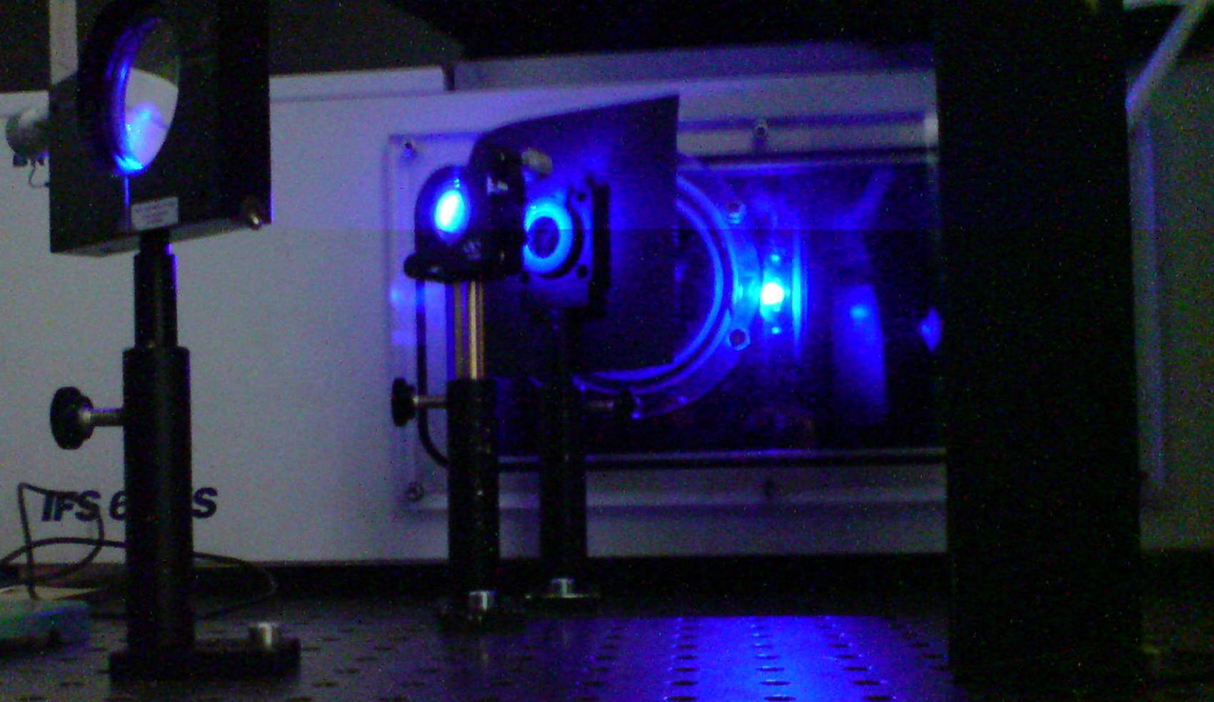
Time-resolved FTIR Spectroscopy
We apply rapid-scan and step-scan techniques to elucidate the mechanism of cyclic processes over a broad time range from 500 nanoseconds to seconds after excitation with a pulsed laser (see Biophys. J. 2009).
The step-scan method covers an important time range, which is not at all or only in principal accessible by ultrafast pump-probe spectroscopy. The recording in step-scan is performed continuously with respect to both frequency and time and thereby leads to a gapless data set.
Data are analyzed by singular value decomposition or by spectrally weighted global fits, which yield kinetic models of the reactions.
We have demonstrated that the time resolution of DC step-scan can be enhanced by suitable electronic synchronization (see JACS 2015).
Method Development
One of our goals is to enable investigation of irreversible reactions and systems with long cycling times:
- by integration of microfluidic flow cells the sample is rapidly exchanged (see PCCP 2013).
- photochemical regeneration in photochromic systems was established in step-scan for the first time (two-color stepscan) (see JACS 2018).
The employment of modern, powerful quantum cascade lasers allows us to enter totally new fields of application. By using frequency combs we record thousands of time-resolved spectra after a single excitation (single shot experiment) (see Anal. Chem. 2018).
We analyze reactions in monolayers with nanometer thickness by ATR spectroscopy (see Langmuir 2018, Langmuir 2019).
We compensate for the lacking spatial information of IR spectroscopy on proteins by double difference spectroscopy:- segment-resolved double difference spectroscopy can assign signals to specific secondary structural elements or domains in proteins (see Biochemistry 2010, Nucleic Acids Res. 2016).
- by isotope labeling and double difference spectroscopy we address single amino acids in a collaboration with theory (see Sci. Rep. 2016).
Artificial amino acids as infrared probes allow us to follow structural changes at specific sites with high time resolution (see PCCP 2019).
Theory
For interpretation and assignment of the infrared spectra, we also perform quantum chemical calculations including explicit models for the environment. Even difference spectra can be calculated (see J. Phys. Chem. Lett. 2010, PCCP 2013).
For very demanding questions and the calculation of double difference spectra we are relying on collaborations (see Sci. Rep. 2016).


- Light Sensors and Optogenetics
Many light responses of animals, plants, fungi and bacteria are governed by the blue region of the sun's spectrum. Examples are the growth of plants towards the light (phototropism) and the setting of the daily rhythm in animals (circadian rhythm).

Blue light photoreceptors are proteins that allow the organism to sense the light conditions in the environment. We are investigating the mechanisms of the signal transfer inside the protein from nanoseconds to minutes using time-resolved vibrational and electronic spectroscopy.
For an overview see Nachr. Chem. 2011 (in German).
All blue light receptors are used as tools in optogenetics to render biological processes artificially light-sensitive. For the development and application of these tools, an understanding of the mechanisms is essential.
Cryptochromes
Cryptochromes are used by all organisms, but strongly differ in function and mechanism. They play a key role in the daily rhythmicity of plants and animals including humans.
Cryptochromes regulate a variety of responses to blue light such as plant development (photomorphogenesis) and the setting of the clock in insects. Even a function as sensor of the magnetic field has been demonstrated.
All cryptochromes contain a derivative of vitamin B2 (flavin adenine dinucleotide) as a chromophore. Our goal is to identify the light-induced processes in cryptochrome by using time-resolved spectroscopy (see JACS 2009, J. Phys. Chem. B 2010, JACS 2012, JACS 2015).
Surprisingly, an animal-like cryptochrome (aCRY) has been identified in green algae, which does not only detect blue but also red light. Therefore, we have identified and characterized the first flavin-containing protein, which is activated by red light (see Plant Cell 2012, J. Biol. Chem. 2017).
Phototropin
Plants use the blue light receptor phototropin to optimize photosynthesis and to prevent harmful exposure to strong irradiation (see Nature 2016).
Phototropin contains two so-called LOV domains that bind a flavin mononucleotide as a light-absorbing molecule. Blue light causes the formation of a covalent linkage between the flavin and the protein. After many seconds, this linkage breaks and thereby the sensor is regenerated.
The steps of the photocycle and its kinetics have been intensively studied by us (see Biophys. J. 2003). Currently, we are dealing with the question of how the signal is transfered inside the protein from sensor to effector (a kinase domain). For our model see Biochemistry 2010.
Aureochrome
Some algae use an aureochrome instead of a phototropin as blue light sensor. Aureochromes contain a sensory LOV domain as well, but the effector is a DNA-binding domain (bZIP domain). Consequently, aureochromes most likely act as light-controlled transcription factors, which is of high interest for biotechnology.
The arrangement of sensor and effector is inverted in comparison to phototropin. We investigate, how a signal can be transfered to the bZIP domain in this inverted arrangement and how thereby the DNA binding is modified (see Biochemistry 2013, Biochemistry 2015, Nucleic Acids Research 2017).


- Biocatalysis for Medicinal Chemistry
Biocatalysis Using Halogenases for Medicinal Chemistry
Important drugs such as antibiotics and cytostatic agents are halogenated compounds. Halogenations can be performed under mild conditions and regioselectively using biocatalysis. The required cofactor, a reduced flavin, can be obtained via chemical regeneration (see ACS Catal. 2019). The goal is to open up new pathways for synthesis of pharmaceutical drugs.
We have established a procedure for light-driven biocatalysis, in which the flavin remains bound to the enzyme. This represents a crucial step in the transformation of halogenases into artificial photoenzymes (see ChemCatChem 2018).
- green chemistry: requires light, air and NaCl
- less formation of unproductive H2O2 by uncoupling reaction
- single-enzyme reaction facilitates engineering
Bioanalysis and Protein Biochemistry
- analytical gel filtration (also under illumination)
- cofactor identification using UV/Vis and fluorescence spectroscopy
- polysaccharide analysis via IR spectroscopy and electrophoresis (FACE)
- quantification of cellular components with IR spectroscopy
For biophysical investigations, we overexpress enzymes and photoreceptors or segments thereof in larger amounts in E. coli (such as the cryptochrome, see J. Biol. Chem. 2007).
- mutagenesis, transformation
- overexpression, cell lysis by French press
- global and selective isotope labeling
- purification via affinity chromatography and gel filtration
- quality control by SDS-PAGE and Western Blot
- phosphorylation assays


- Polymer Materials and Dye Photochemistry
Polymer Materials
FTIR spectroscopy allows us to investigate the structure and composition of organic polymers and other materials (see Angew. Chem. Int. Ed. 2010).
As applications we determine the monomer content in copolymers (see Polymer 2017) and resolve structural changes in thermoresponsive, colloidal particles in water (see PCCP 2018).
As surface-sensitive technique we apply ATR difference spectroscopy on monolayers. By photopolymerization we obtain freestanding 2D nanomembranes with promising properties (see Langmuir 2018, Langmuir 2019).
The employment of quantum cascade lasers as powerful light sources allows us to enter totally new fields of application.
Dye Photochemistry
Organic dyes play an important role as fluorescence markers in spectroscopy and microscopy and as chromophores in sensory proteins and light-driven enzymes. Furthermore they have a great potential for application in the field of photovoltaics and in organic LEDs.
We monitor the reaction steps after excitation with light by time-resolved UV/vis and FTIR spectroscopy. Our aim is to identify intermediates and products as well as to determine the kinetics.
In the focus of our investigations are aqueous media, because water poses special challenges for infrared spectroscopy due to its high intrinsic absorption (see PCCP 2013).
Fluorescence Markers for Super-Resolution Microscopy
The development of super-resolution light microscopy beyond Abbe´s diffraction limit is based on dyes which can be switched off transiently. These non-fluorescent states are often not chemically characterized.
Owing to the high sensitivity of FTIR spectroscopy we can identify the products of these inefficient reations by a combination with quantum chemical calculations. In particular, we can record difference spectra in presence of a very high background signal from reaction partners (see J. Phys. Chem. Lett. 2010).


- Algae as Model Organisms
Impact of Algae in Medicine, Biotechnology, and Climate Change
- Photoreceptors from algae such as channel rhodopsins are applied to artificially activate nerve cells by light (optogenetics).
- The mechanism behind one of the most common human genetic diseases, polycystic kidney disease, and many other diseases has been investigated in the model system Chlamydomonas, because the flagella of the alga have much in common with the primary cilium.
- Algal cells produce hydrogen or isoprene upon illumination and are therefore candidates for a direct, environmentally sound conversion of solar energy into fuel or biomass.
- Algae store about half of the globally available carbon dioxide and are therefore a major factor in the investigation of the climate change.
The Green Alga Chlamydomonas
Chlamydomonas reinhardtii is a unicellular green alga with a size of about 10 micrometers and is found all over the world. It serves as a simple model organism for the investigation of photosynthesis and processing of light signals.
With the help of two flagella it reacts to the light conditions of the environment (phototaxis). It uses a biological clock for daily time keeping, which is synchronized by light (circadian rhythm). To investigate the entrainment of this rhythm Chlamydomonas has already been sent to space on the Space Shuttle.
The Diatom Phaeodactylum
Diatoms are unicellular, photosynthetic organisms, which are found in marine and freshwater environments, for example as part of the plankton. Phaeodactylum tricornutum is one of the best characterized diatoms. Its genome has been fully sequenced.
Diatoms contribute significantly (around one fifth) to primary productivity on Earth. Consequently, one in five oxygen molecules consumed by us has been produced by a diatom.
Photoreceptors
Within the framework of interdisciplinary collaborations established in the DFG Research Group 1261, we are investigating the role and the mechanism of photoreceptors in unicellular model algae.
Typical for a member of the plant family, Chlamydomonas contains phototropin und cryptochromes. However, it is lacking the whole family of the red light-sensitive phytochromes. Instead, the alga contains an animal-like cryptochrome (aCRY), which is the only flavoprotein so far that is activated by red light (see Plant Cell 2012).
Additionally, rhodopsins are found in the green alga such as the channelrhodopsins,
which are responsible for phototaxis. Phototropin protects photosynthesis from harmful light intensities (see Nature 2016). aCRY regulates the reproduction (see Plant Physiol. 2017a) and plant cryptochrome the circadian rhythm (see Plant Physiol. 2017b).The green alga thereby offers the opportunity to study the mechanism of almost all common photoreceptors within a comparatively simple organism.
For our biophysical investigations, we overproduce the photoreceptors or segments thereof in larger amounts in E. coli (such as the cryptochrome, see J. Biol. Chem. 2007). From similarities or differences in the properties of the Chlamydomonas photoreceptors as compared to those of land plants, one can draw conclusions on the relevant processes and observe evolutionary developments.
Phaeodactylum contains different photoreceptors than plants, because it is a secondary endosymbiont. Cryptochromes and phytochromes are still found in the diatom. However, phototropin is not produced. Instead, aureochrome regulates some of the blue light responses such as the acclimation to high light.


