Research

Research Topics
- Time-resolved Spectroscopy
We make use of the fact that the reaction of a chromophore changes characteristically its absorption in the visible range (its color) as well as in the IR range (its normal modes).
FTIR Spectroscopy
Fourier transform infrared (FTIR) spectroscopy allows us to investigate the structure and composition of dyes, proteins, cells, organic polymers and other materials (see Angew. Chem. Int. Ed. 2010).
Advantages compared to UV/Vis spectroscopy are the smaller line width and the large number of transitions resulting in a much higher specificity and a clear identification.
In photosensors, the chromophore reacts with amino acids in the protein environment. These amino acids as well as changes in secondary and tertiary structure can be identified by applying FTIR spectroscopy.
Using light-induced difference spectroscopy, we resolve reactions of chromophores and chemical processes of single amino acids against the background of thousands of normal modes of the complex (protein) environment and of water.
Time-resolved Spectroscopy
Absorption of light by a chromophore initiates a sequence of chemical reactions. Time-resolved spectroscopy is an analytical technique which allows us to elucidate these reactions by identifying intermediates and products and their kinetics.
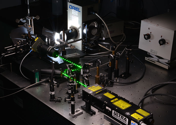
Time-resolved UV/Vis Spectroscopy
In time-resolved UV/Vis spectroscopy we record changes in absorption of white light by the chromophore after excitation with a laser pulse with a duration of a few nanoseconds.
The whole UV/Vis spectrum of the sample is recorded as a difference spectrum at different points in time from 60 nanoseconds to seconds after excitation and analyzed (see Photochem. Photobiol. 2011, J. Biol. Chem. 2025).
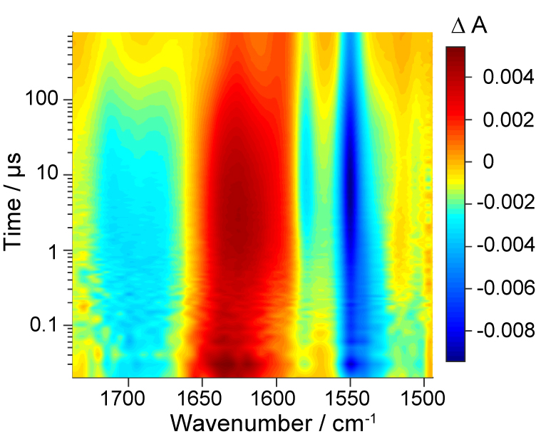
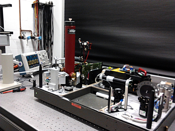
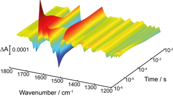
Time-resolved FTIR Spectroscopy
We apply rapid-scan and step-scan techniques to elucidate the mechanism of cyclic processes over a broad time range from 500 nanoseconds to seconds after excitation with a pulsed laser (see Biophys. J. 2009).
The step-scan method covers an important time range, which is not at all or only in principal accessible by ultrafast pump-probe spectroscopy. The recording in step-scan is performed continuously with respect to both frequency and time and thereby leads to a gapless data set.
Data are analyzed by singular value decomposition or by spectrally weighted global fits, which yield kinetic models of the reactions.
We have demonstrated that the time resolution of DC step-scan can be enhanced by suitable electronic synchronization (see JACS 2015).
Method Development
One of our goals is to enable investigation of irreversible reactions and systems with long cycling times:
- by integration of microfluidic flow cells the sample is rapidly exchanged (see PCCP 2013).
- development of a quantum cascade laser setup for nanosecond time resolution and microlitre consumption (see PCCP 2020)
- photochemical regeneration in photochromic systems was established in step-scan for the first time (two-color stepscan) (see JACS 2018).
The employment of modern, powerful quantum cascade lasers allows us to enter totally new fields of application. By using frequency combs we record thousands of time-resolved spectra after a single excitation (single shot experiment) (see Anal. Chem. 2018).
We analyze reactions in monolayers with nanometer thickness by ATR spectroscopy (see Langmuir 2018, Langmuir 2019).
We compensate for the lacking spatial information of IR spectroscopy on proteins by double difference spectroscopy:- segment-resolved double difference spectroscopy can assign signals to specific secondary structural elements or domains in proteins (see Biochemistry 2010, Nucleic Acids Res. 2016).
- by isotope labeling and double difference spectroscopy we address single amino acids in a collaboration with theory (see Sci. Rep. 2016).
Artificial amino acids as infrared probes allow us to follow structural changes at specific sites with high time resolution (see PCCP 2019, PCCP 2021).
Theory
For interpretation and assignment of the infrared spectra, we also perform quantum chemical calculations including explicit models for the environment. Even difference spectra can be calculated (see J. Phys. Chem. Lett. 2010, PCCP 2013, JACS 2025).
For very demanding questions and the calculation of double difference spectra we are relying on collaborations (see Sci. Rep. 2016).

- Spectroscopy in Living Cells
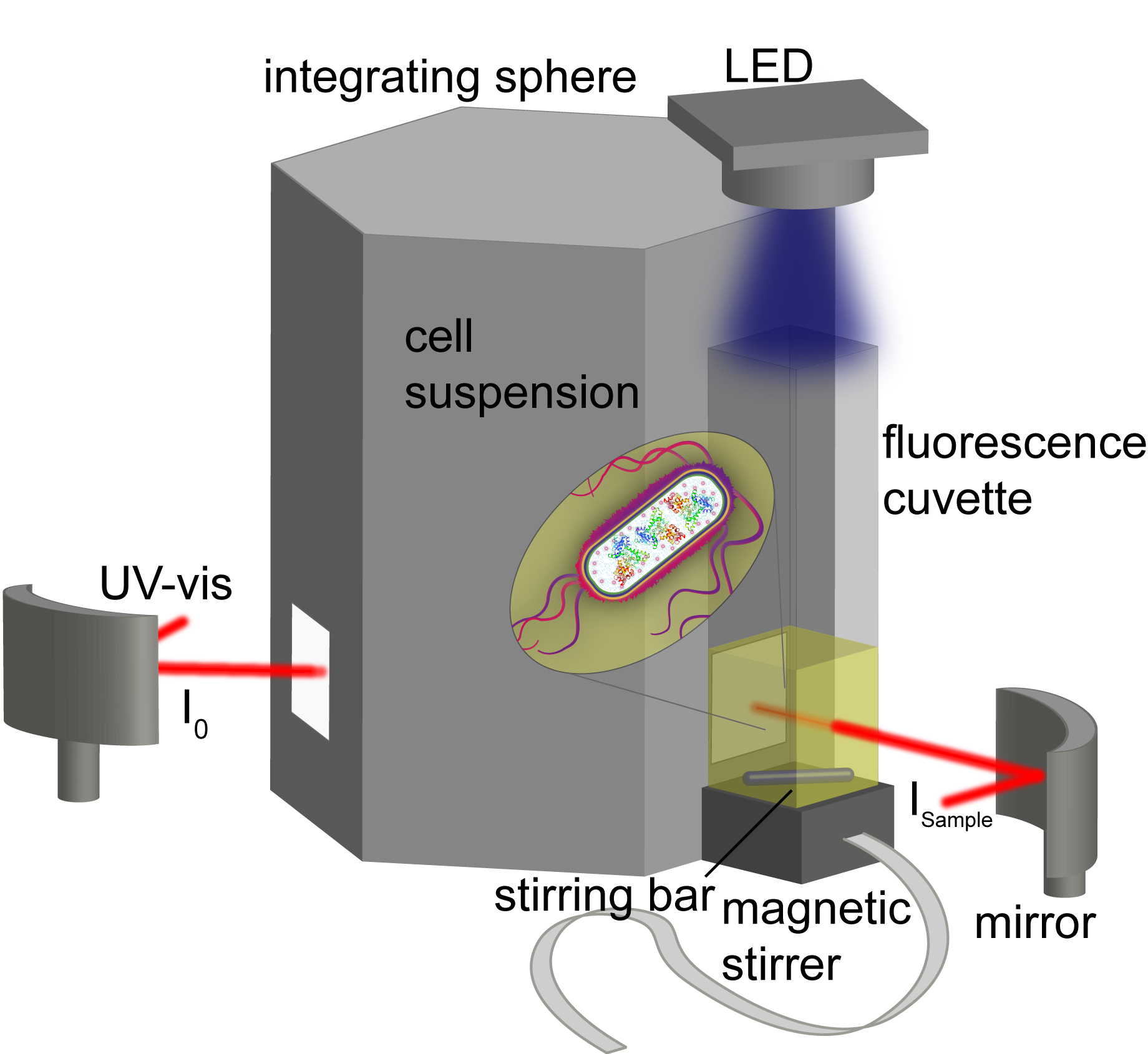
Cellular conditions can strongly impact protein structure and function. The complex cellular environment of proteins cannot be fully emulated in vitro. We study proteins directly in living cells by applying and developing fluorescence, UV-vis and infrared spectroscopy to capture their properties, structures and interactions in a native environment. In addition, in-cell spectroscopy allows us to study proteins that cannot be isolated.
In-Cell Fluorescence and UV-vis Spectroscopy
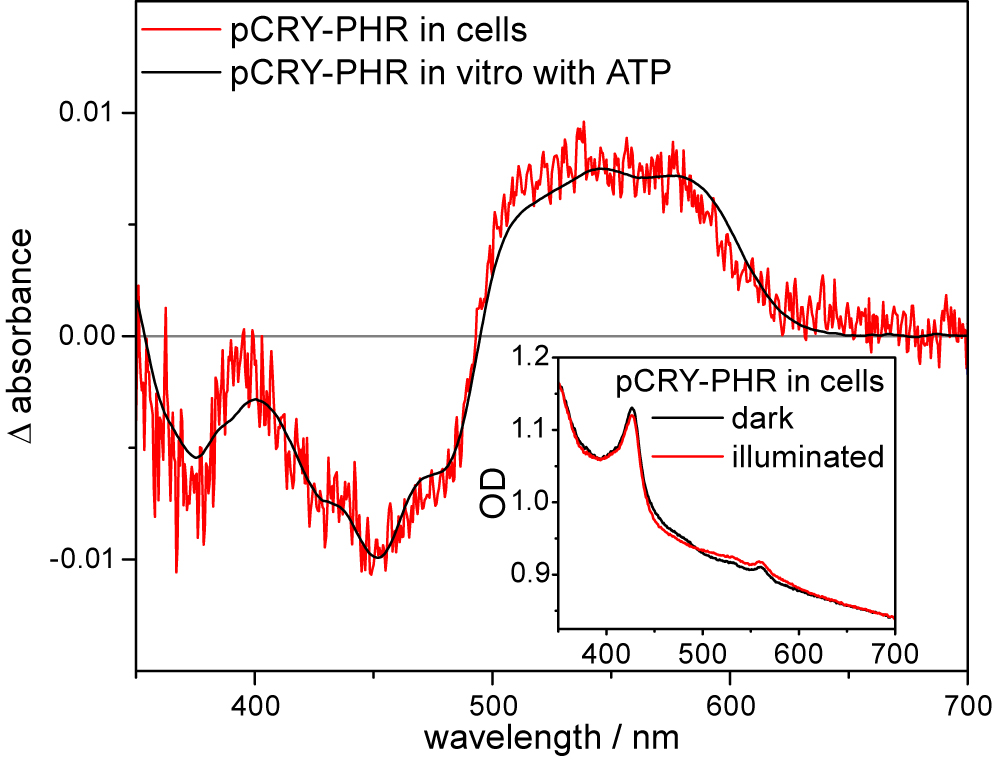
In the visible range, we study changes in absorption and fluorescence of protein cofactors in living cells (see J. Biol. Chem. 2020, J. Phys. Chem. Lett. 2021). The strong scattering by the cells, which challenges UV-vis spectroscopy, is compensated by using an integrating sphere.
We resolve the role of cellular compounds such as ATP in the mechanism of receptors by UV-vis spectroscopy and study kinetics of cofactor conversion in cells by fluorescence spectroscopy (see J. Biol. Chem. 2020).
In-Cell Infrared (FTIR) Spectroscopy
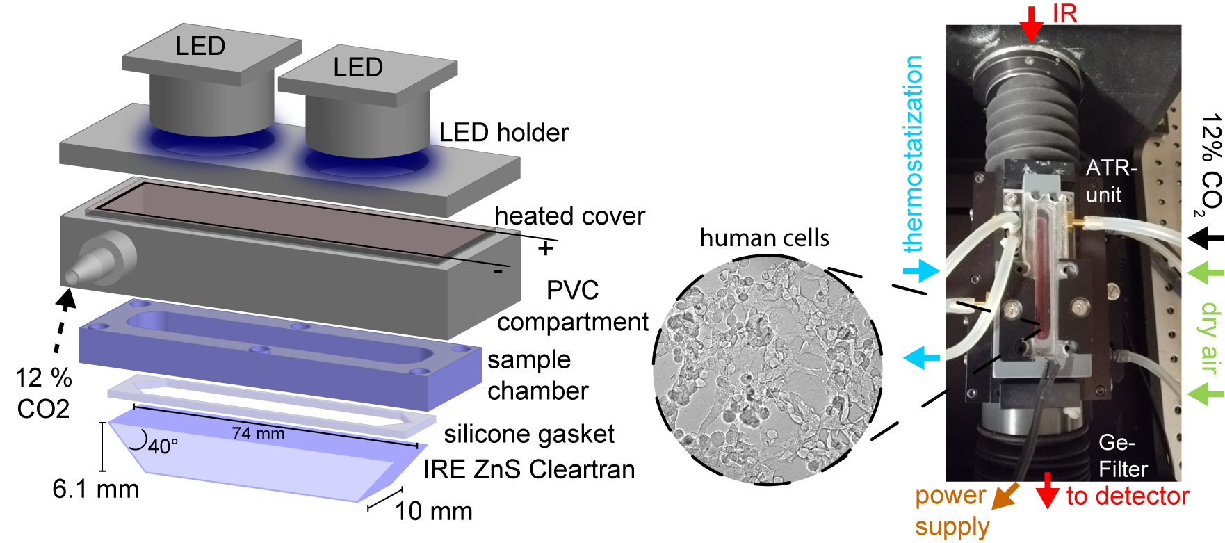
We employ in-cell infrared spectroscopy to quantify cellular components directly within intact cells (see L&O 2018). Further, we have developed in-cell infrared difference spectroscopy (ICIRD) to investigate structural responses of photoreceptors in a native environment (see J. Biol. Chem. 2020). In living bacterial cells, we study receptors with a time resolution of 8 ms using the rapid-scan technique (see Front. Phys. 2023).
Recently, we developed a setup to cultivate, transfect and measure human cell lines for the first time directly inside an infrared spectrometer. This setup allows us to perform ICIRD on receptors in eukaryotic cells (see J. Am. Chem. Soc. 2025).
- Photo-Biocatalysis for Medicinal Chemistry
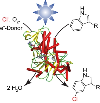
Important drugs such as antibiotics and cytostatic agents are halogenated compounds. Halogenations can be performed under mild conditions and regioselectively using biocatalysis. The required cofactor, a reduced flavin, can be obtained via chemical regeneration (see ACS Catal. 2019). The goal is to open up new pathways for synthesis of pharmaceutical drugs.
Artificial Photoenzymes
We have established a procedure for light-driven biocatalysis, in which the flavin remains bound to the enzyme. This represents a crucial step in the transformation of halogenases into artificial photoenzymes (see ChemCatChem 2018). Studying the catalytic mechanism enables the rational design of halogenase variants with improved photocatalytic properties (see J. Biol. Chem. 2024, Photochem. Photobiol. Sci. 2024).
- green chemistry: requires light, oxygen and NaCl
- less formation of unproductive H2O2 by uncoupling reaction
- single-enzyme reaction facilitates engineering
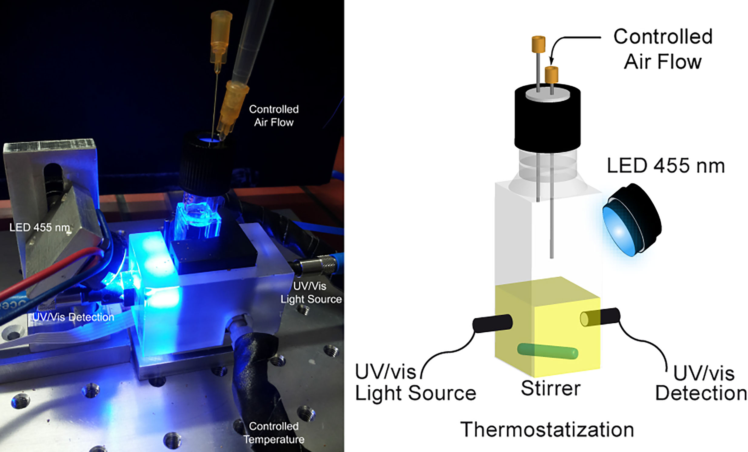
A custom photoreactor was developed for enzymatic photocatalysis, allowing real-time, in situ monitoring of light-driven reactions (see ChemCatChem 2018).
Protein Biochemistry
For biophysical investigations and photocatalysis, we overexpress the enzymes in larger amounts in E. coli:
- mutagenesis, transformation
- overexpression, cell lysis by French press
- global and selective isotope labeling for IR spectroscopy
- purification via FPLC using affinity chromatography, ion exchange and gel filtration
- quality control by SDS-PAGE and Western Blot
Bioanalysis
We apply further methods for the quantitative and qualitative characterization and analysis of enzymes:
- identification and characterization of cofactors using UV/Vis and fluorescence spectroscopy
- determination of dissociation constants via fluorescence anisotropy
- phosphorylation assays
- analytical gel filtration
- Light Sensors and Optogenetics
Many light responses of animals, plants, algae, fungi and bacteria are governed by the blue region of the sun's spectrum. Examples are the the setting of the daily rhythm in animals (circadian rhythm) and the growth of plants towards the light (phototropism).
Blue light photoreceptors are proteins that allow the organism to sense the light conditions in the environment. We are investigating the mechanisms of the signal transfer inside the protein from nanoseconds to minutes using time-resolved vibrational and electronic spectroscopy.
All blue light receptors are applied as tools in optogenetics to render biological processes artificially light-sensitive. For the development and application of these tools, an understanding of the mechanisms is essential.
Cryptochromes
Cryptochromes are used by all organisms, but strongly differ in function and mechanism. They play a key role in the daily rhythmicity of plants and animals including humans.
Cryptochromes regulate a variety of responses to blue light such as plant development (photomorphogenesis) and the setting of the clock in insects. Even a function as sensor of the magnetic field has been demonstrated.
All cryptochromes contain a derivative of vitamin B2 (flavin adenine dinucleotide) as a chromophore. Our goal is to identify the light-induced processes in cryptochrome by using in-cell and time-resolved spectroscopy (see JACS 2009, J. Phys. Chem. B 2010, JACS 2012, JACS 2015, J. Phys. Chem. Lett. 2021).
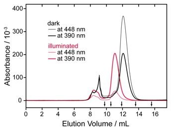
Surprisingly, an animal-like cryptochrome (aCRY) has been identified in green algae, which does not only detect blue but also red light. Therefore, we have identified and characterized the first flavin-containing protein, which is activated by red light (see Plant Cell 2012, J. Biol. Chem. 2017, Biophys. J. 2019).
Phototropin
Plants use the blue light receptor phototropin to optimize photosynthesis and to prevent harmful exposure to strong irradiation (see Nature 2016).
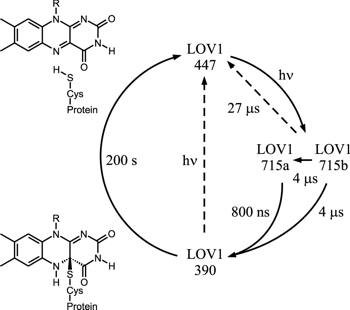
Phototropin contains two so-called LOV domains that bind a flavin mononucleotide as a light-absorbing molecule. Blue light causes the formation of a covalent linkage between the flavin and the protein. After many seconds, this linkage breaks and thereby the sensor is regenerated.
The steps of the photocycle and its kinetics have been intensively studied by us (see Biophys. J. 2003). Currently, we are dealing with the question of how the signal is transfered inside the protein from sensor to effector (a kinase domain). For our model see Biochemistry 2010.
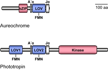
Aureochrome
Some algae use an aureochrome instead of a phototropin as blue light sensor. Aureochromes contain a sensory LOV domain as well, but the effector is a DNA-binding domain (bZIP domain). Consequently, aureochromes most likely act as light-controlled transcription factors, which is of high interest for biotechnology.
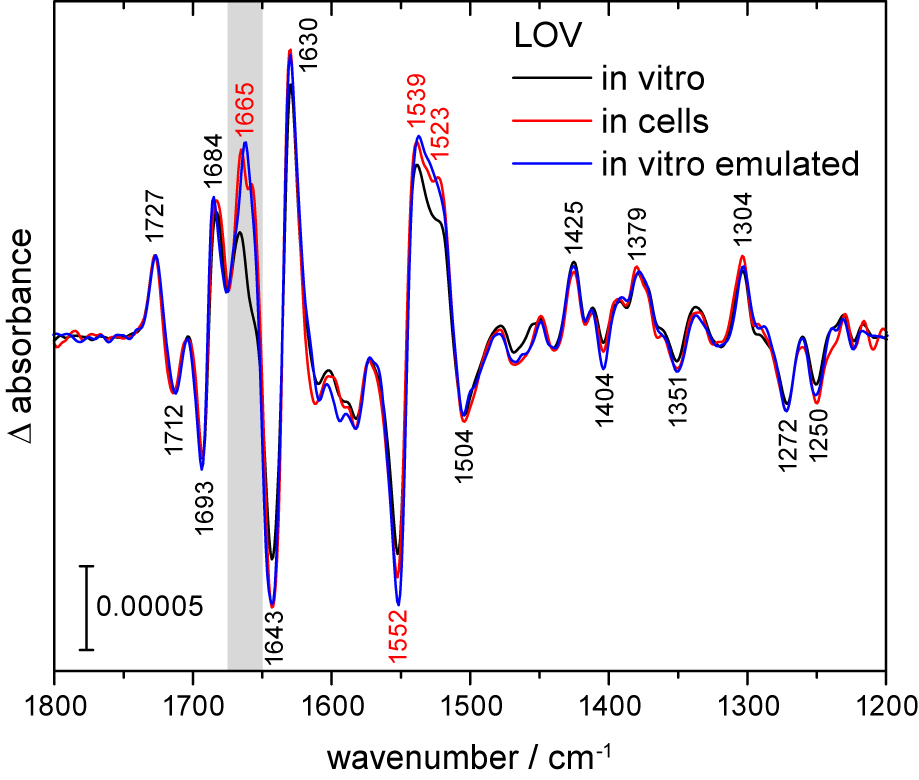
The arrangement of sensor and effector is inverted in comparison to phototropin. We investigate, how a signal can be transfered to the bZIP domain in this inverted arrangement and how thereby the DNA binding is modified (see Biochemistry 2013, Biochemistry 2015, Nucleic Acids Research 2017). In addition, the signal progression from LOV to bZIP is investigated in living cells, to understand the signal progression in a native environment (see JBC 2020, Front. Phys. 2023).
Sample Preparation
We routinely overexpress and isolate photoreceptors (such as cryptochrome, see J. Biol. Chem. 2007) on a large scale in our laboratory. This routine includes mutagenesis, transformation, cell lysis, and various purification techniques such as affinity chromatography, ion exchange chromatography, and gel filtration using fast protein liquid chromatography (FPLC). We perform selective isotope labeling of photoreceptors to assign signals from secondary structure and individual residues (see Photochem. & Photobiol. 2017).

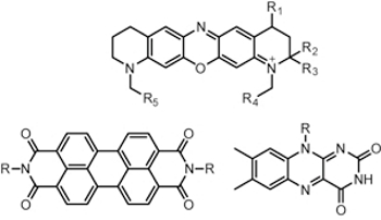
- Oxygen-Dependent Photochemistry in Solution and in Polymer Materials
Dye Photochemistry
Organic dyes play an important role as fluorescence markers in spectroscopy and microscopy and as chromophores in sensory proteins and light-driven enzymes.
Photochemistry in Aqueous Solution
We monitor the reaction steps after excitation with light by time-resolved UV/vis and infrared spectroscopy. Our aim is to identify intermediates and products as well as to determine the kinetics.
In the focus of our investigations are aqueous media, because water poses special challenges for infrared spectroscopy due to its high intrinsic absorption.
Owing to the high sensitivity of FTIR spectroscopy and the high temporal resolution provided by step-scan or quantum cascade lasers, reaction products and intermediates of photochemistry can be identified in combination with quantum chemical calculations.
In particular, we record difference spectra in the presence of a very high background signal from reaction partners in aqueous solution (see J. Phys. Chem. Lett. 2010) and under tightly controlled oxygen concentrations (see PCCP 2013, PCCP 2020).
Polymer Materials
FTIR spectroscopy allows us to investigate the structure and composition of organic polymers and other materials under controlled conditions with high spectral and temporal resolution (see Angew. Chem. Int. Ed. 2010).
As surface-sensitive technique we apply ATR difference spectroscopy on monolayers. By aerobic and anaerobic photopolymerization, we obtain freestanding 2D nanomembranes with promising properties (see Langmuir 2018, Langmuir 2019).
As applications we determine the monomer content in copolymers (see Polymer 2017, Macromol. Chem. Phys. 2021) and resolve structural changes in thermoresponsive, colloidal particles in water (see PCCP 2018, Soft Matter 2022).