© Universität Bielefeld

Super-Resolution Microscopy
A. Investigation of the NF-kB signaling pathway using super-resolution microscopy
In collaboration with Prof. Dr. Mike Heilemann [1, 2], University of Würzburg, we investigate the NF-kB pathway, including the upstream receptors like TNF-R1 using super-resolution microscopy. Super-resolution microscopy is a new technique that improves the spatial resolution of light microscopy to a sub-diffraction resolution up to an order of magnitude per dimension-achieving up to In a current interdisciplinary studies, realized in collaboration with Prof. Heilemann (Würzburg), Prof. Dr. Jean-Baptiste Sibarita and Prof. Dr. Daniel Choquet (Université de Bordeaux, Institut de Neurosciences, 33077 Bordeaux, Cédex, France), we used a variant of single-particle tracking, sptPALM, an approach that exploits the selective photoconversion of fluorescent reporters to resolve the dynamics of dense populations of trafficking molecules with sub-diffraction spatial resolution the mobility of single NF-kB p65 particles in living neurons and of the TNF-R1 in different cell lines [4] [5].
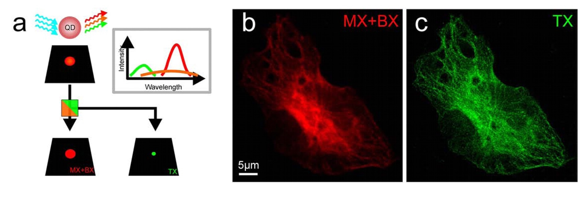
a. QDTI is realized by exciting semiconductor quantum dots QDot655 with 488 nm. As a consequence of multiexciton generation, three excitonic states with different fluorescence emission characteristics are generated. The emission of the triexciton is blue-shifted and can be spectrally separated. As a consequence of the three-photon absorption the resolution is increased ∼2-fold. b, c. Confocal fluorescence images of U373 cells, microtubule network stained by immunofluorescence using secondary antibodies labeled with QDot655. The triexciton channel (c) shows an increase in resolution and less contribution of out-of-focus light (optical sectioning). Figure adapted from [3].
B. Imaging of adult human neural crest derived stem cells by helium-ion microscopy
As described above, light microscopy is usually limited to resolve structures with sizes in the order of the wavelength and above. A few years ago helium-ion microscopes (HIM) became available due to a breakthrough in the development of reliable gas field ionization sources. This type of microscope is very similar to a SEM except it employs helium ions instead of electrons. It posses all the discussed advantages of SEM. However, the edge resolution of HIM was demonstrated to be as small as 2.4 angstrom. Thus it surpasses the currently achievable resolution of TEM for biological specimen. The exceptionally small beam diameter is a consequence of the higher helium ion mass compared to electrons. This leads to a higher momentum at the same energy, i.e. the de Broglie wavelength becomes smaller. Consequently, diffraction related issues are relaxed which allow smaller probe sizes. In addition, further mechanisms improve the achievable image resolution of HIM in comparison to SEM. These are related to differences in the beam-sample-interaction. The helium ion beam broadens substantially less within the first few nanometres of the sample. This is the relevant sample volume where a significant fraction of secondary electrons escapes. A dedicated HIM study concluded that beam broadening “does not currently limit the microscopes image resolution” at all. As the helium ions penetrate deeper into the sample, the beam broadens substantially due to scattering. Some ions are even backscattered to the sample surface which leads to the emission of additional secondary electrons which potentially degrade resolution. However, in HIM the fraction of these secondary electrons to the total amount is negligible. This is very different in SEM. Here the majority of secondary electrons originate from backscattered electrons. Thus these mechanisms lead to the conclusion that HIM still resolves significantly finer surface features on a bulk sample even if the beam size in SEM could be made as small as in HIM.
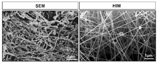
Comparison of coventional scanning electron microscopy (SEM) and helium ion microscopy (HIM) reveals superior spatial resolution of HIM. For SEM fibrin matrix was coated with gold particles. In contrast for HIM imaging no coating is necessary. In a collaboration with Prof. Dr. A. Gölzhäuser and PD. Dr. A.Beyer we were able to use HIM high-resolution analysis for the characterization of a novel three-dimensional fibrin based cell culture system for adult human inferior turbinate stem cells (ITSCs) [6]. Currently, we focus on the monitoring of differentiating ITSCs at different time points after osteogenic, myogenic and neuronal induction. In addition we are interested in HIM-imaging of living differentiating and differentiated ITSCs.
References
[1] M. Heilemann, S. van de Linde, A. Mukherjee, M. Sauer, Super-resolution imaging with small organic fluorophores, Angew Chem Int Ed Engl 48 (2009) 6903-6908.
[2] S. van de Linde, M. Sauer, M. Heilemann, Subdiffraction-resolution fluorescence imaging of proteins in the mitochondrial inner membrane with photoswitchable fluorophores, J Struct Biol 164 (2008) 250-254.
[3] M. Heidbreder, U. Endesfelder, S. van de Linde, S. Hennig, D. Widera, B. Kaltschmidt, C. Kaltschmidt, M. Heilemann, Subdiffraction fluorescence imaging of biomolecular structure and distributions with quantum dots, Biochim Biophys Acta 1803 (2010) 1224-1229.
[4] C. Zander, T. Engelen, D. Widera, D. Nair, M. Heidbreder, J.B. Sibarita, D. Choquet, M. Heilemann, B. Kaltschmidt, C. Kaltschmidt, Retrograde, dynein-mediated transport of NF-kappaB p65 in hippocampal neurons occurs via heterocomplex formation with HSP90/Hsc70 protein chaperones, submitted for publication (2011).
[5] M. Heidbreder, C. Zander, S. Malkusch, D. Widera, B. Kaltschmidt, C. Kaltschmidt, D. Nair, D. Choquet, J.B. Sibarita, M. Heilemann, TNF-alpha influences the lateral dynamics of TNF receptor I in living cells, Submitted for publication (2011).
[6] J. Greiner, S. Hauser, D. Widera, F. Qunneis, J. Müller, C. Zander, I. Martin, J. Mallah, C. Prante, H. Schwarze, W. Prohaska, A. Beyer, K. Rott, A. Hütten, A. Gölzhäuser, H. Sudhoff, C. Kaltschmidt, B. Kaltschmidt, Efficient animal-serum free 3D cultivation method for adult human neural crest-derived stem cell therapeutics, in revision (2011).