Cell Biology

Further Reading Material
- NF-κB in the Nervous System
Physiological effects of NF-κB within the nervous system
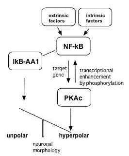
In the nervous system NF-κB is a transcription factor regulated by various extracellular stimuli such as neurotransmitters (glutamate), growth factors (NGF), proinflammatory cytokines (TNF) and others. We discovered that NF-κB is constitutively activate in mature glutamatergic neurons through continuous neurotransmission. In this line NF-κB is necessary for the normal physiological function of a neuron, that is survival and information processing/memory formation. NF-κB controls a neuroprotective gene expression program, which can protect neurons against various toxic substances such as A-beta peptides typically for Alzheimer's disease (Fig. 1).
Future Projects
Under which conditions is NF-κB neuroprotective? Which molecular pathways are influenced by NF-κB in memory formation? How does NF-κB control neuronal circuits and neurogenesis in the adult aging hippocampus?
To study these questions we are using primary neuronal cultures and transgenic mouse models in which neuronal/glial NF-κB is repressed as well as approaches from systems biology.Transport of NF-κB in neurons
We found that NF-κB could be transported retrogradely back from the synapse to the nucleus. We study which motor proteins might be involved in this transport and what might be the physiological function. We are using live imaging, proteomic techniques and super resolution light microscopy (sptPALM).
Neural Stem Cells
We are interested in signals which stimulate proliferation and differentiation of neural stem cells as well as the integration of newborn neurons into neuronal networks. We use several experimental paradigms: neurosphere cultures as well as transgenic mouse models. Moreover we could isolate neural stem cells from various sources including human tissues. We aim to develop the human stem cells as a cellular therapy for craniofacial lesions and neurodegenerative diseases (Fig. 2).
- NF-κB in Stem Cells
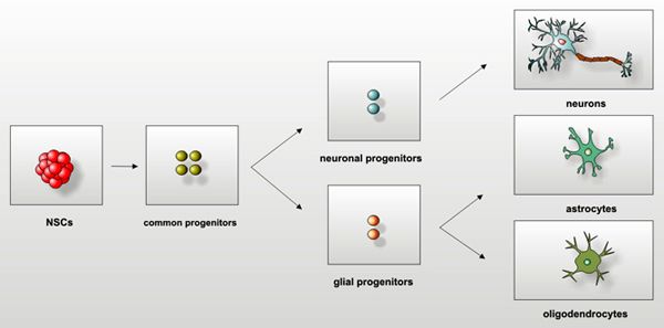
During the development of the mammalian central nervous system (CNS), multipotent precursor cells (stem cells) undergo division, cell fate specification, and maturation. These neural stem cells (NSCs) are characterized by the ability to proliferate and to differentiate into multiple cell types, e.g. neurons or glial cells (Fig. 3).
Embryonic and adult neural stem cells perform self-renewal or differentiate into more committed progenitor cells. These common progenitors give rise to glial and neuronal precursors, which can terminally differentiate into neurons and glial cells. In contrast to neural stem cells, neuronal progenitors give rise to neuronal and not to glial progeny.
In pathological situations, such as brain inflammation, NSCs have been described to ameliorate the disease either due to increased proliferation and replacement of the degenerated tissue or due to secretion of protective cytokines. Much of the inflammatory signal transduction can be considered as the innate immune response triggered by tumor necrosis factor (TNF), one of the crucial inflammation mediators and a well described activator of the transcription factor Nuclear Factor-kappaB (NF-κB).
We were able to demonstrate, that NF-κB and its target genes play a pivotal role in the regulation of neural stem cells biology. In particular, NF-κB regulates the TNF mediated proliferation of neural stem cells [1]. In in vitro cultures of NSC, TNF-α mediated NF-κB activation is responsible for fast re-aggregation of neurosphere cultures [2]. Importantly, a deregulation of the pathway leads to tumorigenic transformation of NSCs [3].
Furthermore we described for the first time, that MCP-1, a well known NF-κB target, efficiently regulates the migration of NSCs [4].
Currently, we are interested in the role of NF-κB in various aspects of the stem cell biology - proliferation, differentiation, migration and potential transformation of 'normal' stem cells into cancerous phenotype (reviewed in [5, 6, 7]).
References
[1] D. Widera, I. Mikenberg, M. Elvers, C. Kaltschmidt, B. Kaltschmidt, Tumor necrosis factor alpha triggers proliferation of adult neural stem cells via IKK/NF-κB signaling, BMC Neurosci 7 (2006) 64.
[2] D. Widera, I. Mikenberg, A. Kaus, C. Kaltschmidt, B. Kaltschmidt, Nuclear Factor-kappaB controls the reaggregation of 3D neurosphere cultures in vitro, Eur Cell Mater 11 (2006) 76-84; discussion 85.
[3] A. Kaus, D. Widera, S. Kassmer, J. Peter, K. Zaenker, C. Kaltschmidt, B. Kaltschmidt, Neural stem cells adopt tumorigenic properties by constitutively activated NF-κB and subsequent VEGF up-regulation, Stem Cells Dev 19 (2010) 999-1015.
[4] D. Widera, W. Holtkamp, F. Entschladen, B. Niggemann, K. Zanker, B. Kaltschmidt, C. Kaltschmidt, MCP-1 induces migration of adult neural stem cells, Eur J Cell Biol 83 (2004) 381-387.
[5] D. Widera, C. Kaltschmidt, B. Kaltschmidt, NF-κB, Potential Role in Adult Neural Stem Cells, in: Hirokawa, Windhorst, Hirsch (Eds.), Encyclopedia of Neuroscience, Springer Verlag, Berlin, 2009, pp. 2862 - 2867.
[6] D. Widera, I. Mikenberg, B. Kaltschmidt, C. Kaltschmidt, Potential role of NF-κB in adult neural stem cells: the underrated steersman?, Int J Dev Neurosci 24 (2006) 91-102.
[7] D. Widera, A. Kaus, C. Kaltschmidt, B. Kaltschmidt, Neural stem cells, inflammation and NF-κB: basic principle of maintenance and repair or origin of brain tumours?, J Cell Mol Med 12 (2008) 459-470.
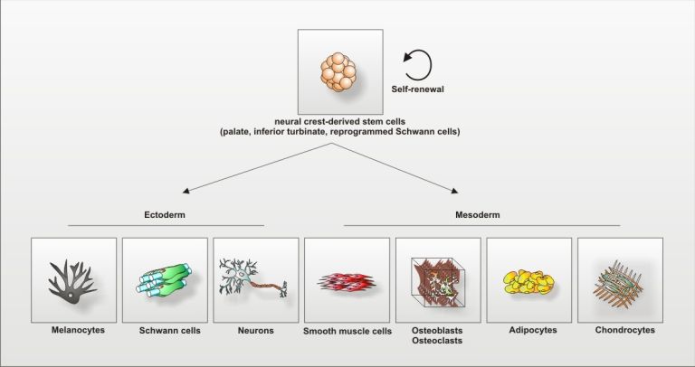
- Neural Crest-derived Stem Cells
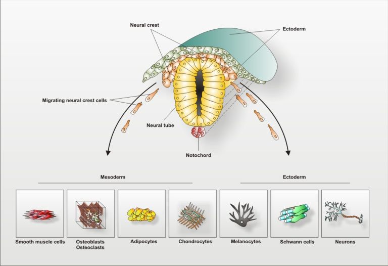
The so called Neural Crest was firstly described by 1868 by Wilhem His as the 'Zwischenstrang', the Intermediate Chord, as it appeared between the Neural Chord and the future ectoderm in the development of the chick embryo. After neurulation, neural crest cells migrate out to give rise to various mesodermal and ectodermal cell populations such as peripheral neurons, cartilage forming chondrocytes of bone-forming osteoblasts (see Fig. 4).
Recently, the persistence of neural crest-related stem cells was reported in the adult [8, 9, 10]. Such neural crest-derived stem cells (NCSCs) could exhibit a dormant stem cell population in the adulthood and have the capacity for both,- self-renewal and generation of multiple progenies in vitro and in vivo (see Fig. 5).
In our laboratory we discovered such NCSCs within the human periodontal ligament [8], rodent and human palate [9] and within the respiratory mucosa of the human inferior nasal turbinate [10].
Furthermore, we identified adult Schwann cells as a further population of neural-crest-related cells harboring stem cell properties [11]. We demonstrated, that such cells can be easily reprogrammed to multipotency by culture alone.
Currently, we focus on the molecular mechanisms regulating stem cell properties of NCSCs concerning especially the role of the transcription factor NF-κB.Our work on NCSCs is supported by the grant 01GN1006A of the German Ministry of Education and Research (BMBF).
References
[8] D. Widera, W.D. Grimm, J.M. Moebius, I. Mikenberg, C. Piechaczek, G. Gassmann, N.A. Wolff, F. Thevenod, C. Kaltschmidt, B. Kaltschmidt, Highly efficient neural differentiation of human somatic stem cells, isolated by minimally invasive periodontal surgery, Stem Cells Dev 16 (2007) 447-460.
[9] D. Widera, C. Zander, M. Heidbreder, Y. Kasperek, T. Noll, O. Seitz, B. Saldamli, H. Sudhoff, R. Sader, C. Kaltschmidt, B. Kaltschmidt, Adult palatum as a novel source of neural crest-related stem cells, Stem Cells 27 (2009) 1899-1910.
[10] S. Hauser, D. Widera, F. Qunneis, J. Müller, C. Zander, J. Greiner, C. Strauss, P. Lüningschrör, P. Heimann, H. Schwarze, J. Ebmeyer, H. Sudhoff, M.J. Araúzo-Bravo, B. Greber, H. Zaehres, H. Schöler, C. Kaltschmidt, B. Kaltschmidt, Isolation of Novel Multipotent Neural Crest-Derived Stem Cells from Adult Human Inferior Turbinate, Stem Cells and Developement - in revision (2011).
[11] D. Widera, P. Heimann, C. Zander, Y. Imielski, M. Heidbreder, M. Heilemann, C. Kaltschmidt, B. Kaltschmidt, Schwann Cells Can Be Reprogrammed to Multipotency by Culture, Stem Cells Dev (2011).
- Subunit-specific Role of NF-κB in Cancer
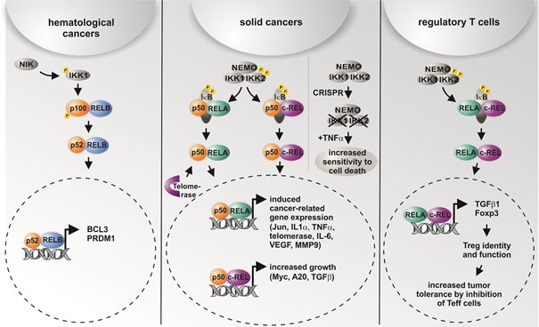
The transcription factor NF-κB is a key player in inflammation, cancer development, and progression. NF-κB stimulates cell proliferation, prevents apoptosis, and could promote tumor angiogenesis as well as metastasis. Extending the commonly accepted role of NF-κB in cancer formation and progression, we summarize in a recently published review that different NF-κB subunits have been shown to be active and of particular importance in distinct types of cancer [12]. Here, overexpression data gained by database mining argue against a universal mechanism of cancer-mediated activation of NF-κB, and suggest a much more elaborated mode of NF-κB regulation, indicating a tumor type-specific upregulation of the NF-κB subunits. While non-canonical NF-κB RELB signaling is described to be mostly present in hematological cancers, solid cancers reveal constitutive canonical NF-κB RELA or c-REL activity (see Fig. 6). Based on these findings, our current research aims to understand the distinct roles of NF-κB subunits and their upstream kinases in cancer cells in more detail. On methodical level, we apply CRISPR/Cas-mediated knockout strategies for guided deletions of NF-κB-subunits in human cells. In this regard, we recently observed TNF-mediated cell death only in human cells lacking IKK1 and IKK2 and not in single CRISPR/Cas-mediated IKK knockouts, suggesting that both IKK1 and IKK2 are required for functional TNF-signaling [13]. We further demonstrated a knockout of c-REL in human in human cervical cancer cells to result in significantly reduced proliferative behavior and a significant delay in the prometaphase of mitosis. Compared to the wild type, an increased resistance against chemotherapeutic agents was observable in c-REL knockout cells [14], emphasizing direct clinical implications of NF-κB-signaling for the development of new treatment strategies.
References
[12] Kaltschmidt B., Greiner J.F.W., Kadhim, H.M. & Kaltschmidt, C. 2018. Subunit-Specific Role of NF-kB in Cancer. Biomedicines.
[13] Slotta, C., Storm, J., Pfisterer, N., Henkel, E., Kleinwachter, S., Pieper, M., Ruiz-Perera, L. M., Greiner, J. F. W., Kaltschmidt, B. & Kaltschmidt, C. 2018. IKK1/2 protect human cells from TNF-mediated RIPK1-dependent apoptosis in an NF-kappaB-independent manner. Biochim Biophys Acta.
[14] Slotta, C., Schulter T., Ruiz-Perera, L. M., Kadhim, H. M., Tertel, T., Henkel, E., Hubner, W., Greiner, J. F. W., Huser, T., Kaltschmidt, B. & Kaltschmidt, C. 2017. CRISPR/Cas9-mediated knockout of c-REL in HeLa cells results in profound defects of the cell cycle. PLoS One, 12, e0182373.
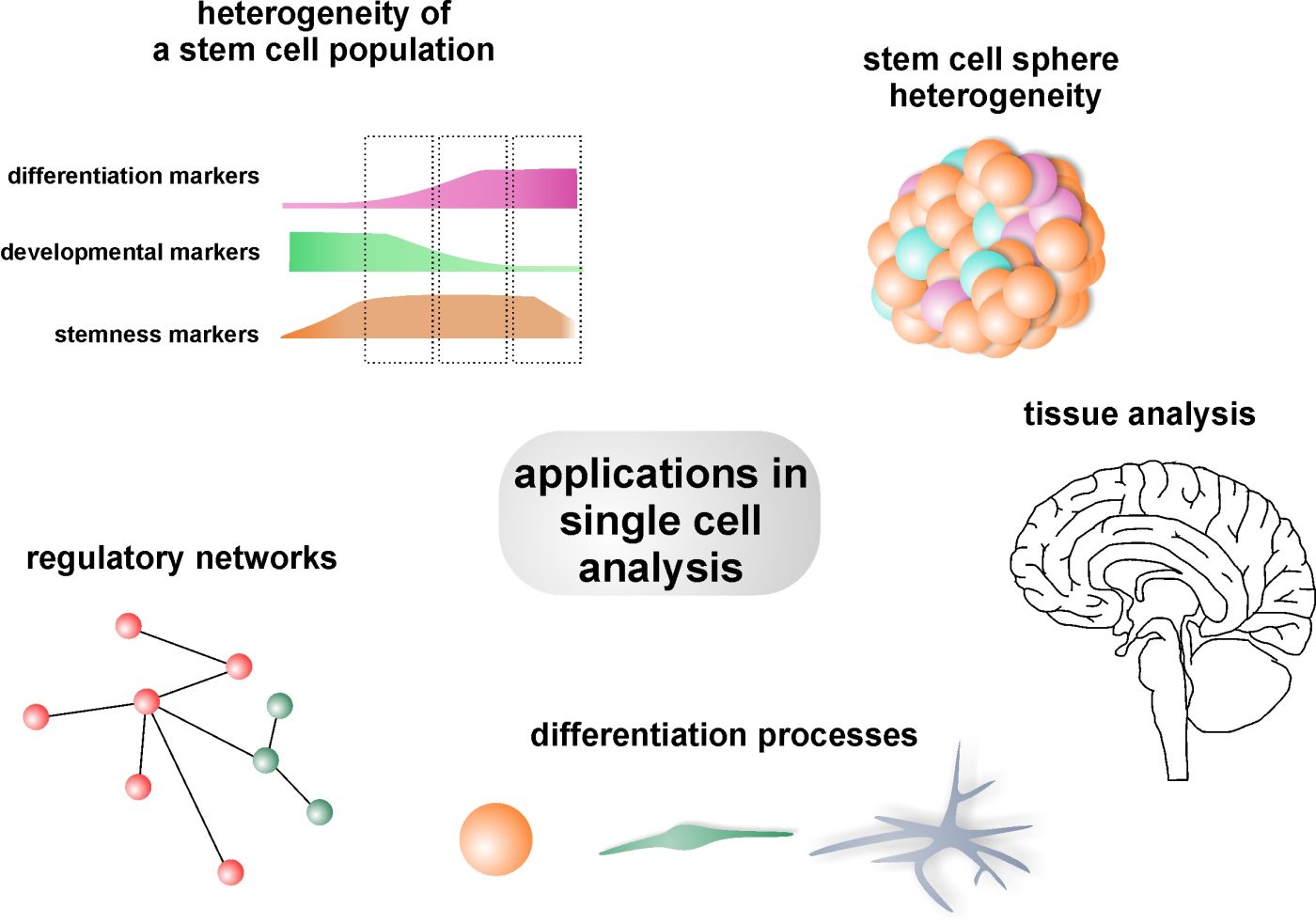
- Single Cell Analysis
Understanding lineage choices of stem cells in context of development and differentiation is commonly assessed using bulk samples masking cellular heterogeneity and dynamics. Addressing this challenge, profiling of individual cells serves a rapidly developing state-of-the arte technique to more precisely define stem cell populations as well as differentiated and intermediate cell types with great implications for understanding development and disease progression. Among the huge variety of adult human stem cells, neural crest derived inferior turbinate stem cells (ITSCs) reveal a remarkably high differentiation potential into neuro-ectodermal an mesodermal cell-types. Nestin-positive ITSCs can be easily isolated from the inferior turbinate of the human nasal cavity and were reported to be capable of functionally recovering a PD rat model, showing their great regenerative potential in vivo.
To successfully analyse single ITSCs and nuclei, we apply flow cytometric fluorescence activated cell sorting (FACS) followed by molecular biological analyses like transcriptional profiling using SMARTSeq2 and RNA-Seq. We are particularly interested in comparing transcriptional data of single ITSCs in the context of differentiation into neuronal cell types and mesodermal derivates. Single cell profiling methods will allow us to investigate cell-to-cell variability during differentiation processes of adult human neural crest derived stem cells for addressing developmental questions and designing accurate disease models. Figure 7 shows different applications for single cell analysis.
- Biomechanical Properties of Cells
It is known that the mechanical properties of a cell give insight into the state the cell is e.g. health or state of differentiation.
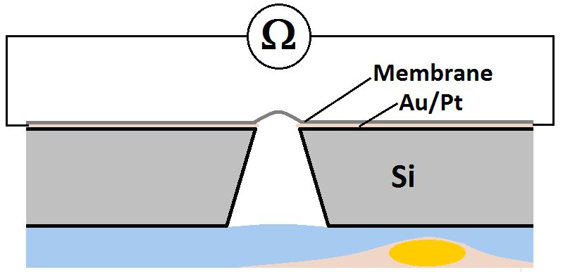
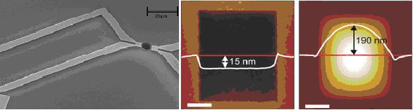
Within an interdisciplinary project we try to make a complete new approach to investigate the mechanical properties of a single cell. Our goal is to build a microscopic microphone as sketched below in Fig. 8.The basis of the microphone is a 1nm thick Carbon Nanomembrane (CNM) made out of a cross linked aromatic self-assembled monolayer in the workgroup of Prof Dr. Armin Golzhäuser (Uni Bielefeld). This membrane is highly flexible and furthermore shows interesting piezoresistive properties. This renders the membrane an ideal tool to receive weak sound waves and transform the sound into a measurable voltage drop.
To accomplish this, the membrane is immobilized on a silicon chip, which is custom-made in the working team of Dr. Thomas Weimann at the Physikalisch-Technische Bundesanstalt. This chip serves primary as a retainer, but also enables the efficient electrical contacting of the CNM (Fig. 9).With the help of this microphone it is/should be possible to detect weak sound waves emitted by a single cell, without inducing big amount of mechanical stress into the cell. These sound waves might be a passive echo-like reaction to an external sound signal or even have their origin in the internal processes of a living cell.
- Super-Resolution Microscopy
A. Investigation of the NF-kB signaling pathway using super-resolution microscopy
In collaboration with Prof. Dr. Mike Heilemann [15, 16], University of Würzburg, we investigate the NF-κB pathway, including the upstream receptors like TNF-R1 using super-resolution microscopy. Super-resolution microscopy is a new technique that improves the spatial resolution of light microscopy to a sub-diffraction resolution up to an order of magnitude per dimension-achieving up to In a current interdisciplinary studies, realized in collaboration with Prof. Heilemann (Würzburg), Prof. Dr. Jean-Baptiste Sibarita and Prof. Dr. Daniel Choquet (Université de Bordeaux, Institut de Neurosciences, 33077 Bordeaux, Cédex, France), we used a variant of single-particle tracking, sptPALM, an approach that exploits the selective photoconversion of fluorescent reporters to resolve the dynamics of dense populations of trafficking molecules with sub-diffraction spatial resolution the mobility of single NF-κB p65 particles in living neurons and of the TNF-R1 in different cell lines (Fig. 10) [18].
B. Imaging of adult human neural crest derived stem cells by helium-ion microscopy
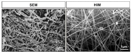
As described above, light microscopy is usually limited to resolve structures with sizes in the order of the wavelength and above. A few years ago helium-ion microscopes (HIM) became available due to a breakthrough in the development of reliable gas field ionization sources. This type of microscope is very similar to a SEM except it employs helium ions instead of electrons. It posses all the discussed advantages of SEM. However, the edge resolution of HIM was demonstrated to be as small as 2.4 angstrom. Thus it surpasses the currently achievable resolution of TEM for biological specimen. The exceptionally small beam diameter is a consequence of the higher helium ion mass compared to electrons. This leads to a higher momentum at the same energy, i.e. the de Broglie wavelength becomes smaller. Consequently, diffraction related issues are relaxed which allow smaller probe sizes. In addition, further mechanisms improve the achievable image resolution of HIM in comparison to SEM. These are related to differences in the beam-sample-interaction. The helium ion beam broadens substantially less within the first few nanometres of the sample. This is the relevant sample volume where a significant fraction of secondary electrons escapes. A dedicated HIM study concluded that beam broadening “does not currently limit the microscopes image resolution” at all. As the helium ions penetrate deeper into the sample, the beam broadens substantially due to scattering. Some ions are even backscattered to the sample surface which leads to the emission of additional secondary electrons which potentially degrade resolution. However, in HIM the fraction of these secondary electrons to the total amount is negligible. This is very different in SEM. Here the majority of secondary electrons originate from backscattered electrons. Thus these mechanisms lead to the conclusion that HIM still resolves significantly finer surface features on a bulk sample even if the beam size in SEM could be made as small as in HIM (Fig. 11).
Comparison of coventional scanning electron microscopy (SEM) and helium ion microscopy (HIM) reveals superior spatial resolution of HIM. For SEM fibrin matrix was coated with gold particles. In contrast for HIM imaging no coating is necessary. In a collaboration with Prof. Dr. A. Gölzhäuser and PD. Dr. A.Beyer we were able to use HIM high-resolution analysis for the characterization of a novel three-dimensional fibrin based cell culture system for adult human inferior turbinate stem cells (ITSCs) [20]. Currently, we focus on the monitoring of differentiating ITSCs at different time points after osteogenic, myogenic and neuronal induction. In addition we are interested in HIM-imaging of living differentiating and differentiated ITSCs.
References
[15] M. Heilemann, S. van de Linde, A. Mukherjee, M. Sauer, Super-resolution imaging with small organic fluorophores, Angew Chem Int Ed Engl 48 (2009) 6903-6908.
[16] S. van de Linde, M. Sauer, M. Heilemann, Subdiffraction-resolution fluorescence imaging of proteins in the mitochondrial inner membrane with photoswitchable fluorophores, J Struct Biol 164 (2008) 250-254.
[17] M. Heidbreder, U. Endesfelder, S. van de Linde, S. Hennig, D. Widera, B. Kaltschmidt, C. Kaltschmidt, M. Heilemann, Subdiffraction fluorescence imaging of biomolecular structure and distributions with quantum dots, Biochim Biophys Acta 1803 (2010) 1224-1229.
[18] C. Zander, T. Engelen, D. Widera, D. Nair, M. Heidbreder, J.B. Sibarita, D. Choquet, M. Heilemann, B. Kaltschmidt, C. Kaltschmidt, Retrograde, dynein-mediated transport of NF-kappaB p65 in hippocampal neurons occurs via heterocomplex formation with HSP90/Hsc70 protein chaperones, submitted for publication (2011).
[19] M. Heidbreder, C. Zander, S. Malkusch, D. Widera, B. Kaltschmidt, C. Kaltschmidt, D. Nair, D. Choquet, J.B. Sibarita, M. Heilemann, TNF-alpha influences the lateral dynamics of TNF receptor I in living cells, Submitted for publication (2011).
[20] J. Greiner, S. Hauser, D. Widera, F. Qunneis, J. Müller, C. Zander, I. Martin, J. Mallah, C. Prante, H. Schwarze, W. Prohaska, A. Beyer, K. Rott, A. Hütten, A. Gölzhäuser, H. Sudhoff, C. Kaltschmidt, B. Kaltschmidt, Efficient animal-serum free 3D cultivation method for adult human neural crest-derived stem cell therapeutics, in revision (2011).- Bioinformatics of the NF-κB-System
The aim of the project is to explore and reconstruct the biological NF-κB transcription factor signal transduction system for better understanding. In particular we plan to map out the mechanisms that convert a chemical stimulus to a cell into a specific cellular response in the example of neural stem cells and adult neurons in mice.
Due to the complexity of pathway interactions and large numbers of components involved in cell proliferation, cell differentiation, signal transduction, cellular rhythms, and cell-to-cell communication, it is quite difficult to intuitively understand the behavior of cellular networks. Recent experimental and computational progress yields networks of increased size and complexity that need to be examined carefully.
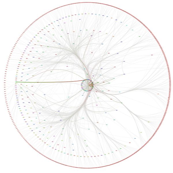
A common way to access the information in a network is through direct visualization, but this fails as it often only results in “fur balls” from which little insight can be gathered. Therefore we discover a new visualization approach, to highlight and communicate one particular piece of information about dynamic network structures. Moreover, we endeavor to find a lossless transformation of dynamic signaling networks into a compact, less redundant representation. We are investigating novel representations of networks, which reduce network complexity by explicitly representing re-occurring network motifs and dynamics, without any loss of information.
As a first result, we present a new approach to network visualization that tightly integrates network analysis methods and edge bundling techniques. The approach uses edge centrality measures to drive a force directed edge bundling method; this results in pictures that clearly show the most significant topological skeleton structures of the input network. We also introduce a new force-directed radial layout that shows group analysis of the k-cores: this results in pictures that show the important cohesive subgroup structures of the input network and their relationships (see Fig. 12).
The foundational modeling ideas and the relationship between network structure and system dynamics is a rapidly expanding frontier of this new science. Of particular interest is the understanding of the organization, complexity and dynamics of biological networks and how these are influenced by network evolution and functionality.
We aim at developing a theoretical framework for the function of specific signal integration modules in the local and global network context. Theories are in developing to address specific aspects of the network function, ranging from, for example, (1) graphical cell-based models to describe development and (2) network construction based on data and text mining to (3) quantitative models with implemented reaction kinetics and mass action relationships to describe information flow.
In order to reconstruct and analyze the NF-κB system we have begun to make use of data mining and information fusion by integrating databases with different contents: e.g. interacting partners and target genes. For the most important elements within the system we have generated experimental and database generated network systems that are regulated by the NF-κB transcription factor. The collected data and information is a combination of tightly interlinked complex systems at various levels of magnitudes.
Each network is constructed using a few basic mechanistic motifs/modules. The functionality of each network implicates participation of specific interacting proteins, where some proteins execute few, other many interactions. The networks operate in the dimension of time and network performance in agreement with cell requirements demands regulation of its activity, e.g. by feedback mechanisms to enable reliable cell fate decisions. Network regulation depends on additional interactions that are clearly visible in so called multidimensional networks.
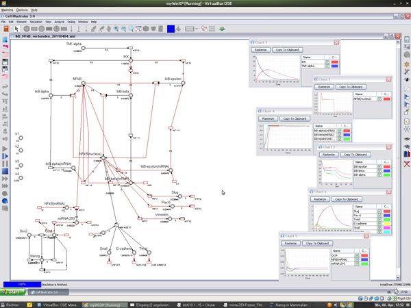
The constructed models are examples of how the behavior of cells modeled by a Petri net can be simulated (see Figure 13).
Ideas for laboratory experiments can be gained and tested by changing the basic Petri net. One approach is a theoretical knockout experiment. For this task, knocked out genes are modeled by deleting the corresponding transitions. A change of gene sequences influencing the catalytic efficiency can be modeled by changing speed parameters of the corresponding transition. Factors outside the cells that influence the signal molecules, the diffusion speed of the medium, and the availability of nutrients or even processes inside the cell can also be modeled.