Light Microscopy Technology Platform of Bielefeld University

Instruments
Confocal Microscopes
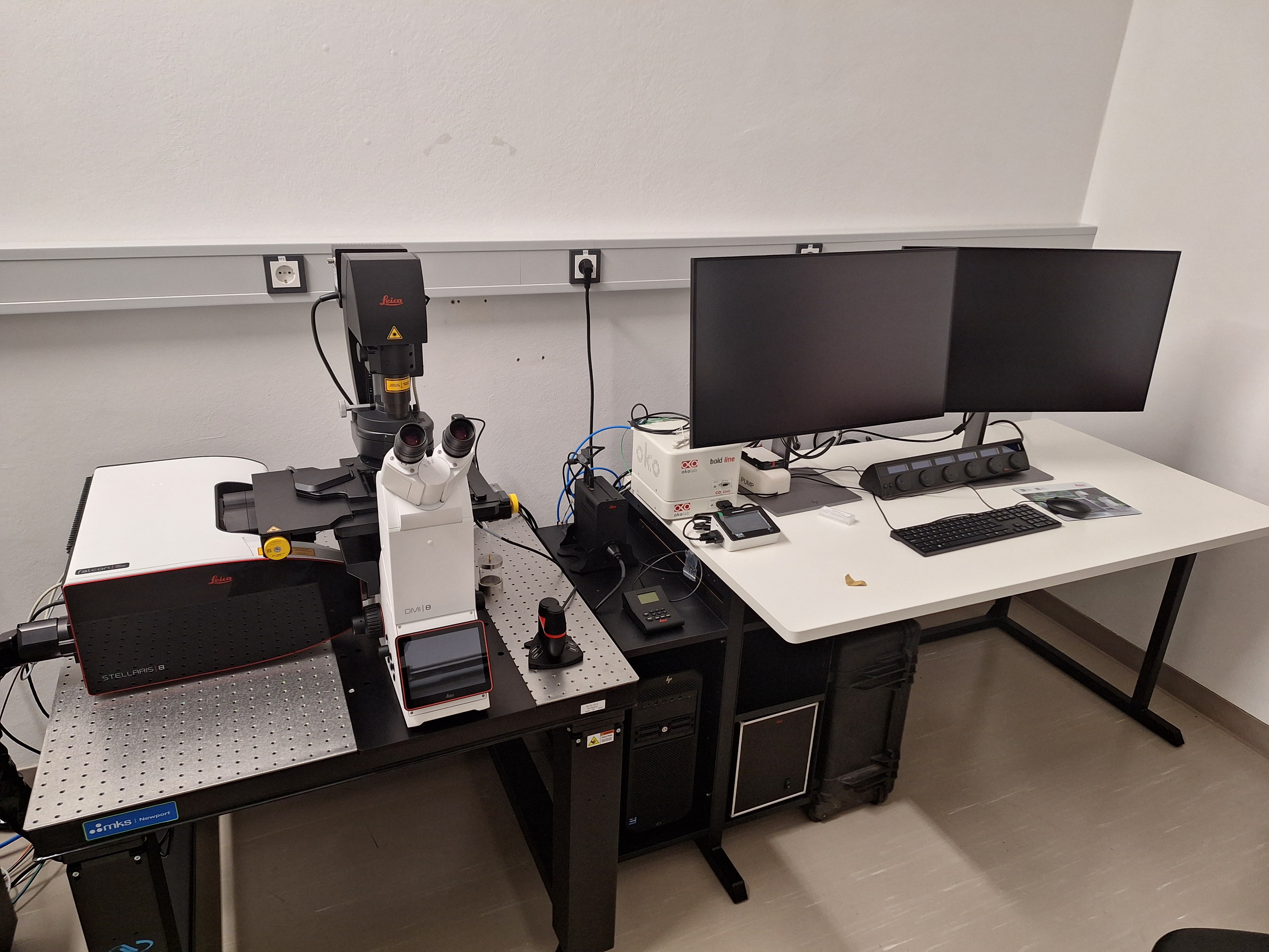
- Leica STELLARIS 8 FALCON
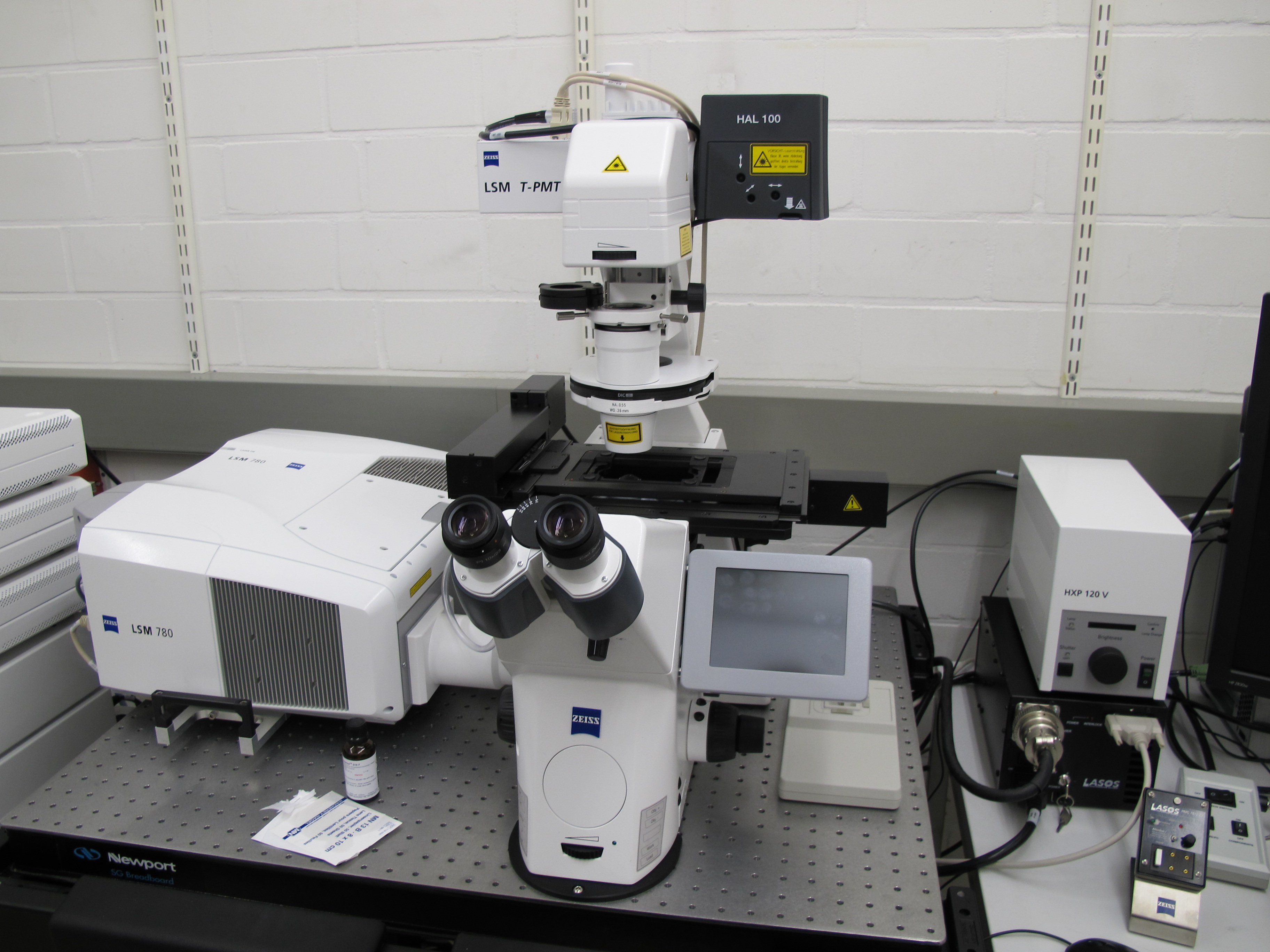
- Zeiss LSM 780
Inverted confocal laser scanning microscope & FCS (W01)
- Laser lines: 405 nm, 458 nm, 488 nm, 514 nm, 561 nm, 633 nm
- Detectors: 2 photomultipliers, 32-ch GaAsp-detector
- Optional: temperature-controlled stage with CO2-incubation
- Software: Zeiss ZEN 2011
Phone at microscope: 5615
Contact: Thorsten Seidel
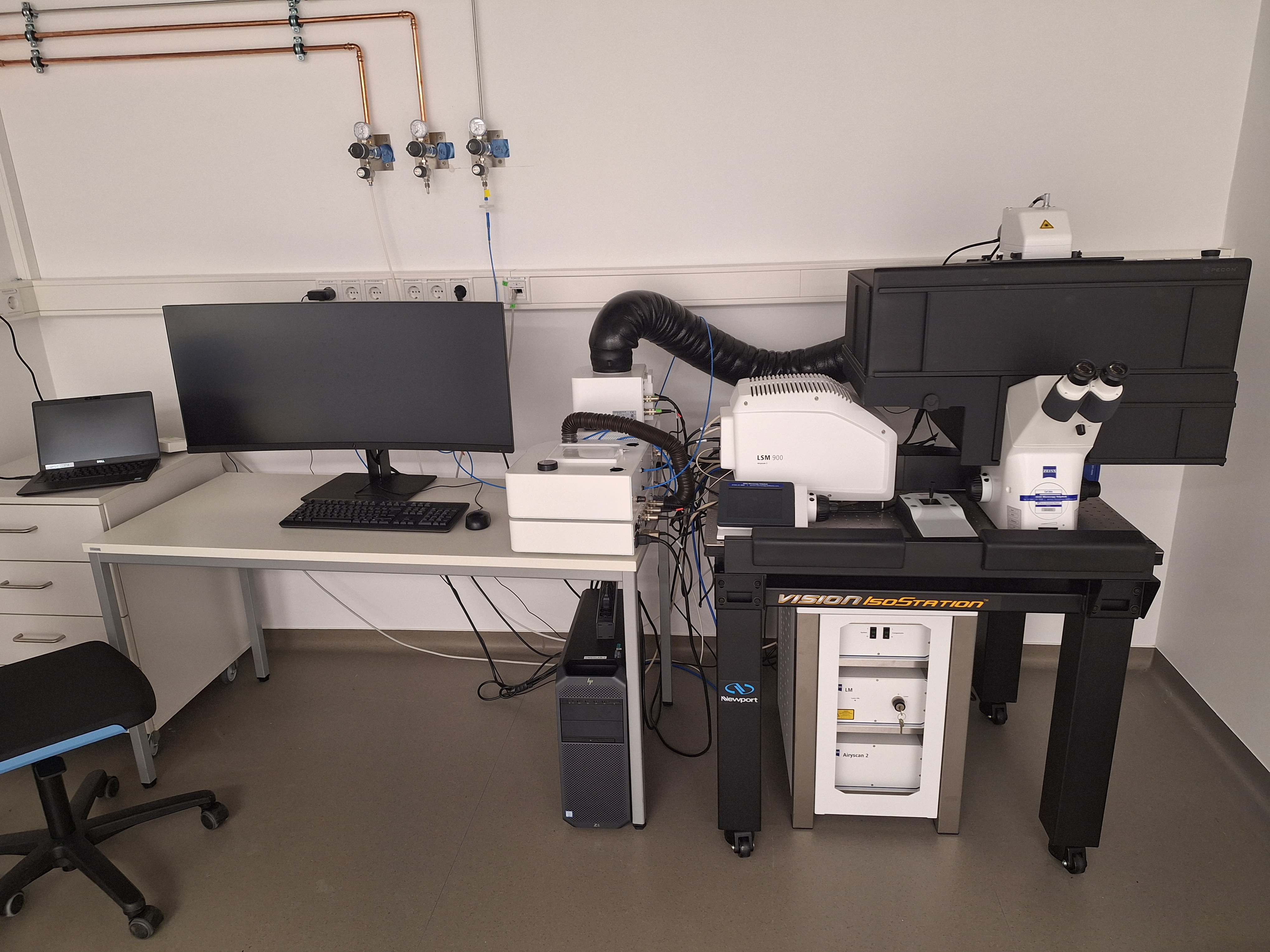
- Zeiss LSM 900
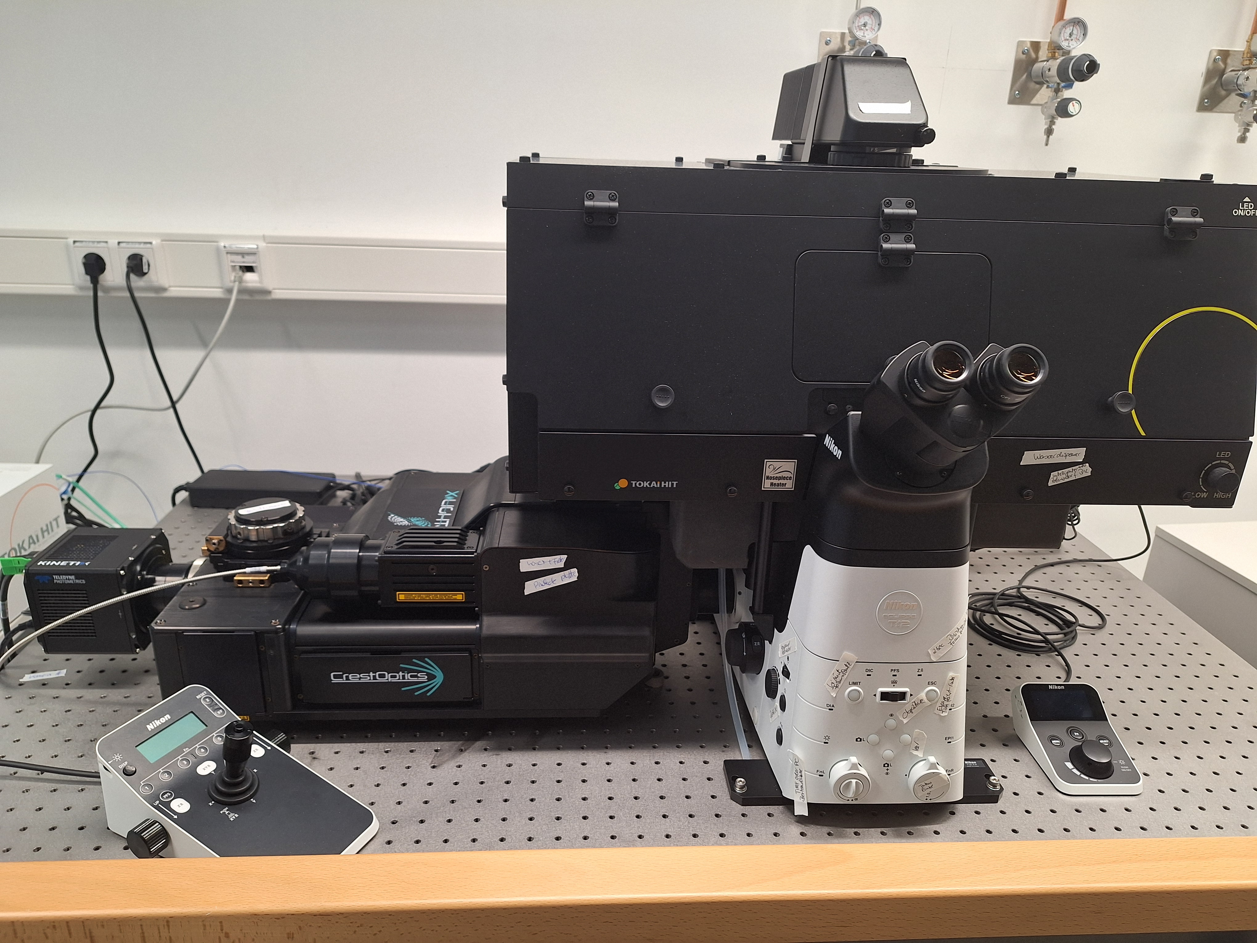
- Spinning Disc Nikon Ti2E Crest X-Light V3
Inverted Stand Nikon Ti2E with Crest Spinning Disc (Building R1-D2)
- Objectives: 10x/0.45, 20x/0.8, 40x/0.95, 40x/1.15, 60x/1.42
- Laser lines: 405 nm, 446 nm, 476 nm, 518 nm, 546 nm, 637 nm, 748 nm
- Camera: Photometrix Kinetics
- Temperature-controlled with CO2-incubation
- Nikon NIS Elements
Phone at microscope: xxxx
Contact: Barbara Biermann
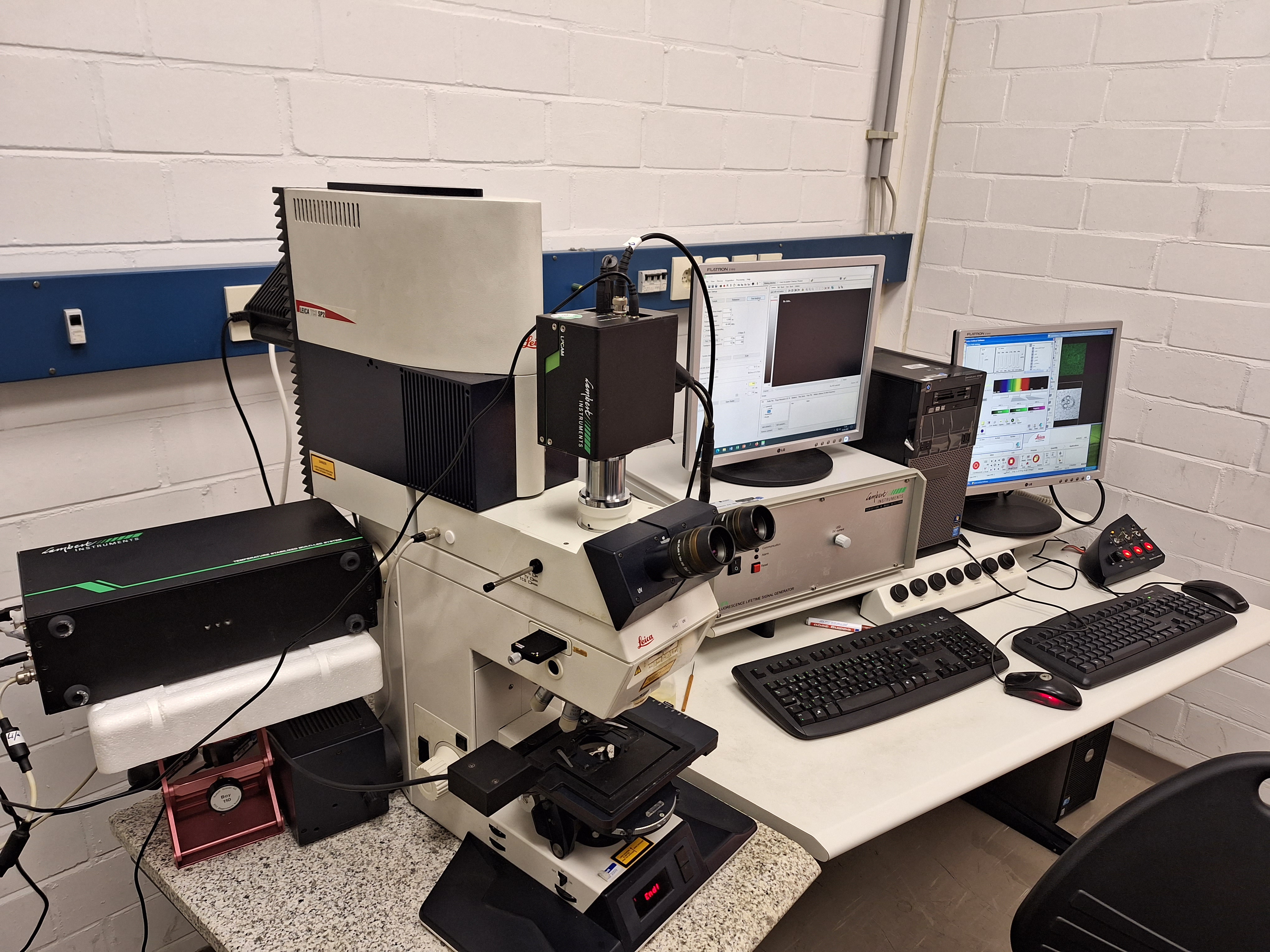
- Leica SP2 mit LIFA FLIM
Upright confocal laser scanning microscope (W1)
- Laser lines: 458 nm, 476 nm, 488 nm, 514 nm, 543 nm, 633 nm
- Filtersets: DD458/514, DD488/543, TD488/543/633, RSP500
- Detectors: 3 photomultipliers
- Optional: temperature-controlled stage
- Software: Leica LCS
Upright stand FLIM (frequency domain)
Lambert LIFA with multi-LED (485 nm, 540 nm, 635 nm)
Filtersets for Fluorescein (long pass), Rhodamin (long pass) and GFP (short pass)
Phone at microscope: 12706
Contact: Thorsten Seidel
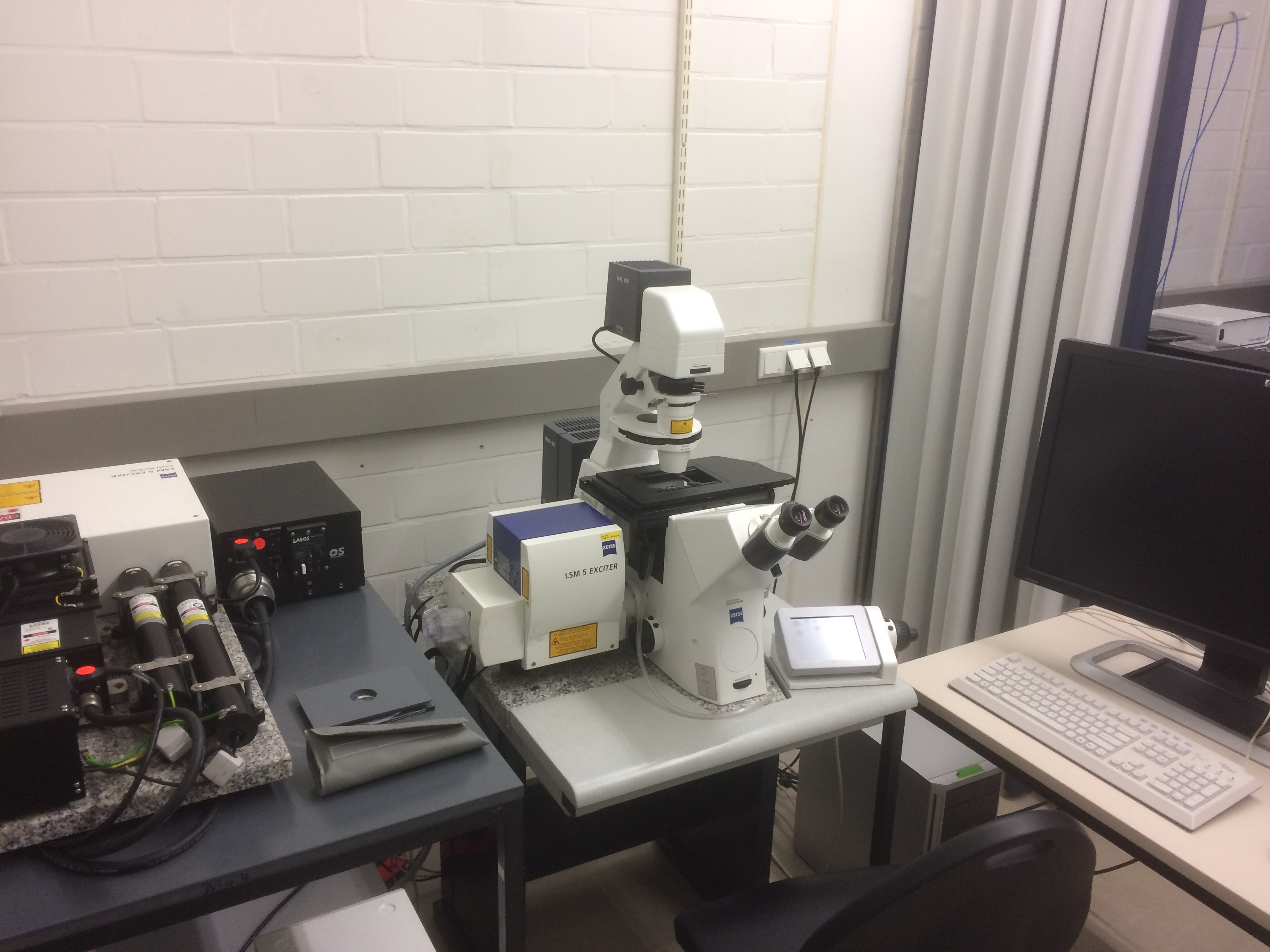
- Zeiss LSM 5 Exciter
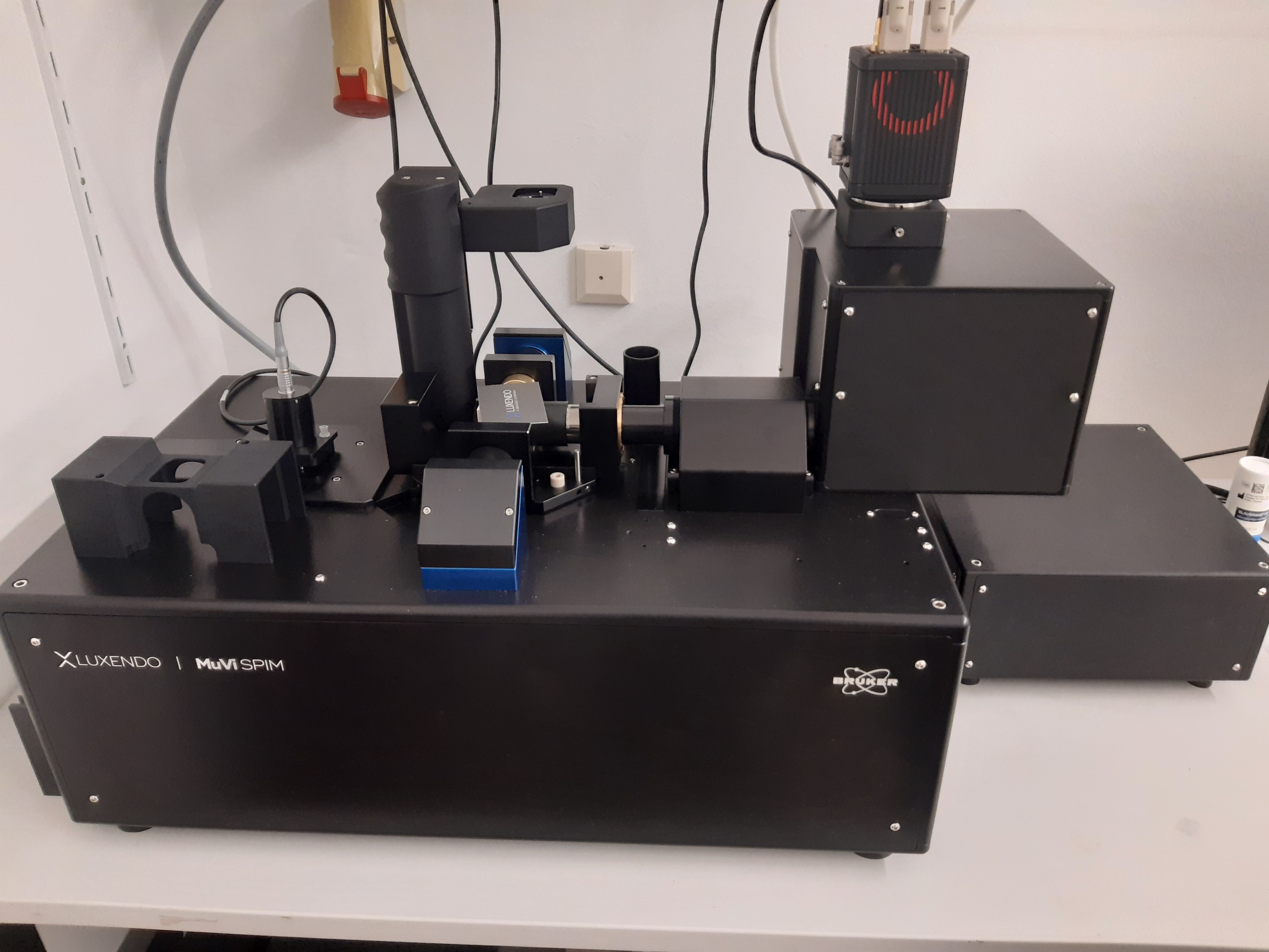
Light Sheet Microscope
Fluorescence Widefield Microscopes
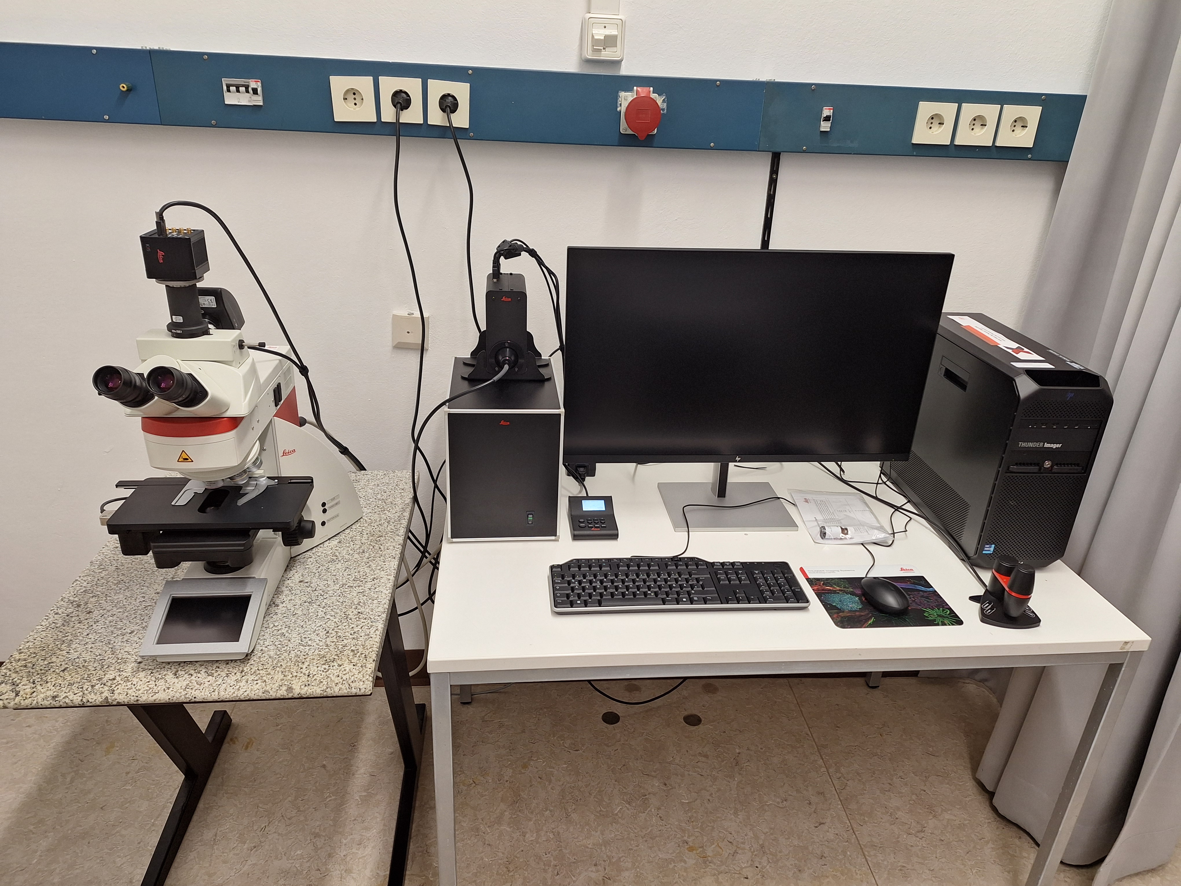
- Leica Thunder Imager Tissue
Leica DM6B motorized upright fluorescence microscope (W0-220)
- Light source: coolLED pE300
- Objectives: 5x/0.15, 10x/0.32 Ph, 20x/0.55, 40x/0.80, 100x/1.32 Oil
- Filter sets: DAPI, GFP, Texas Red, Cy5
- Cameras: Leica K5 and Leica K3C
- Software: Leica LAS X with Thunder module
Phone at microscope: 67437
Contact: Thorsten Seidel
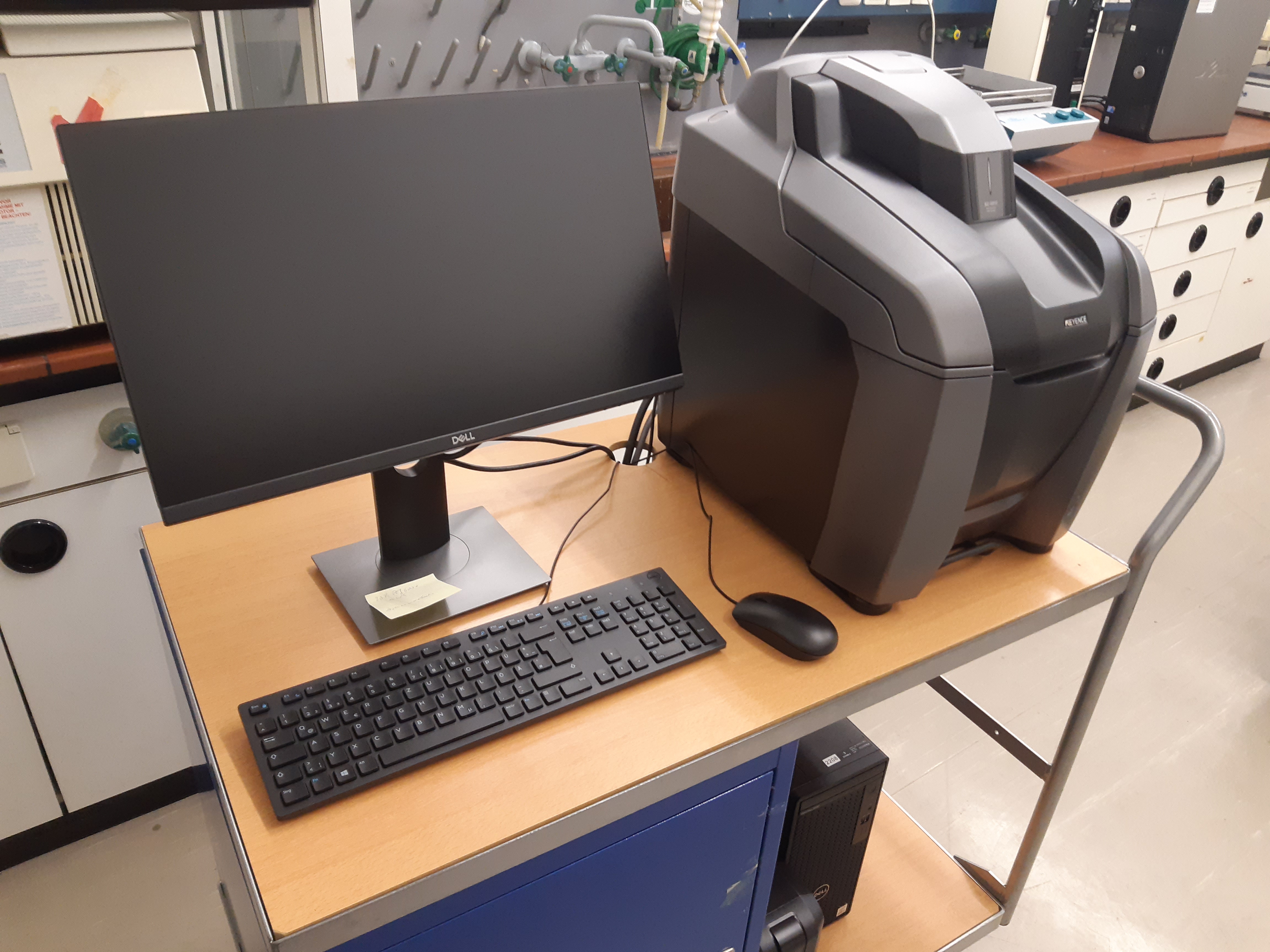
- Keyence BZ-X800
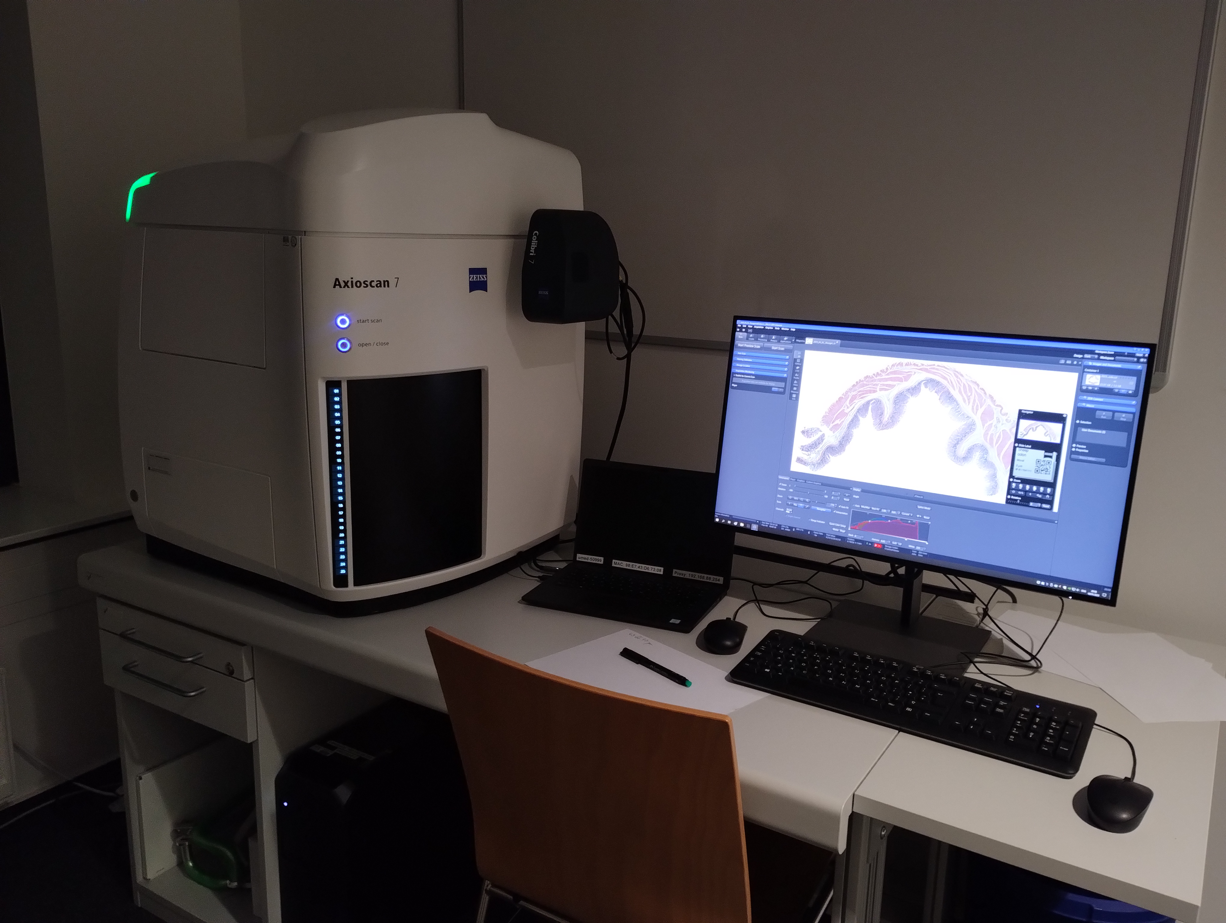
- Zeiss Axioscan
Microscopes, Polarization
Polarization Microscopy allows for visualization of birefringent structures such as cell walls, starch, spindles.
- Octax PolScope
Polarization microscope (W0-211)
- Inverted stand: Nikon Eclipse TE2000-S
- Contrast methods: transmitted light (polarisator & analysator)
- Software: Octax Eyeware
- Camera: Octax camera system
- Applications: imaging of birefringerant structures (spindles, cell walls)
Phone at microscope: 67436
Contact: Thorsten Seidel
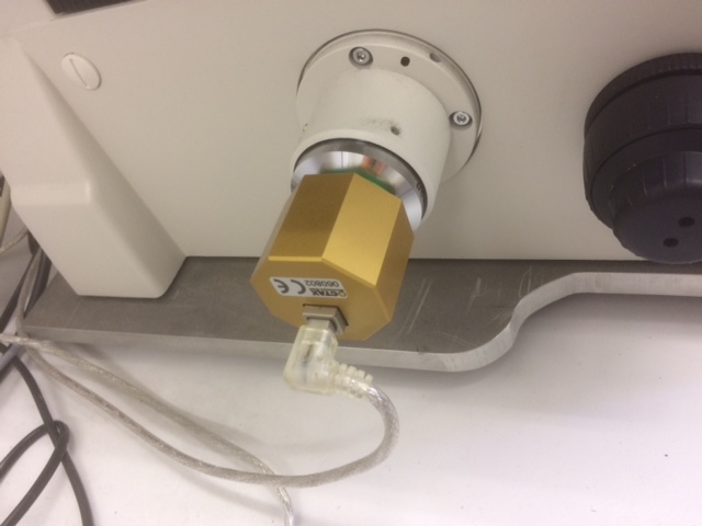
Micro-Manipulation Setup
Other Equipment
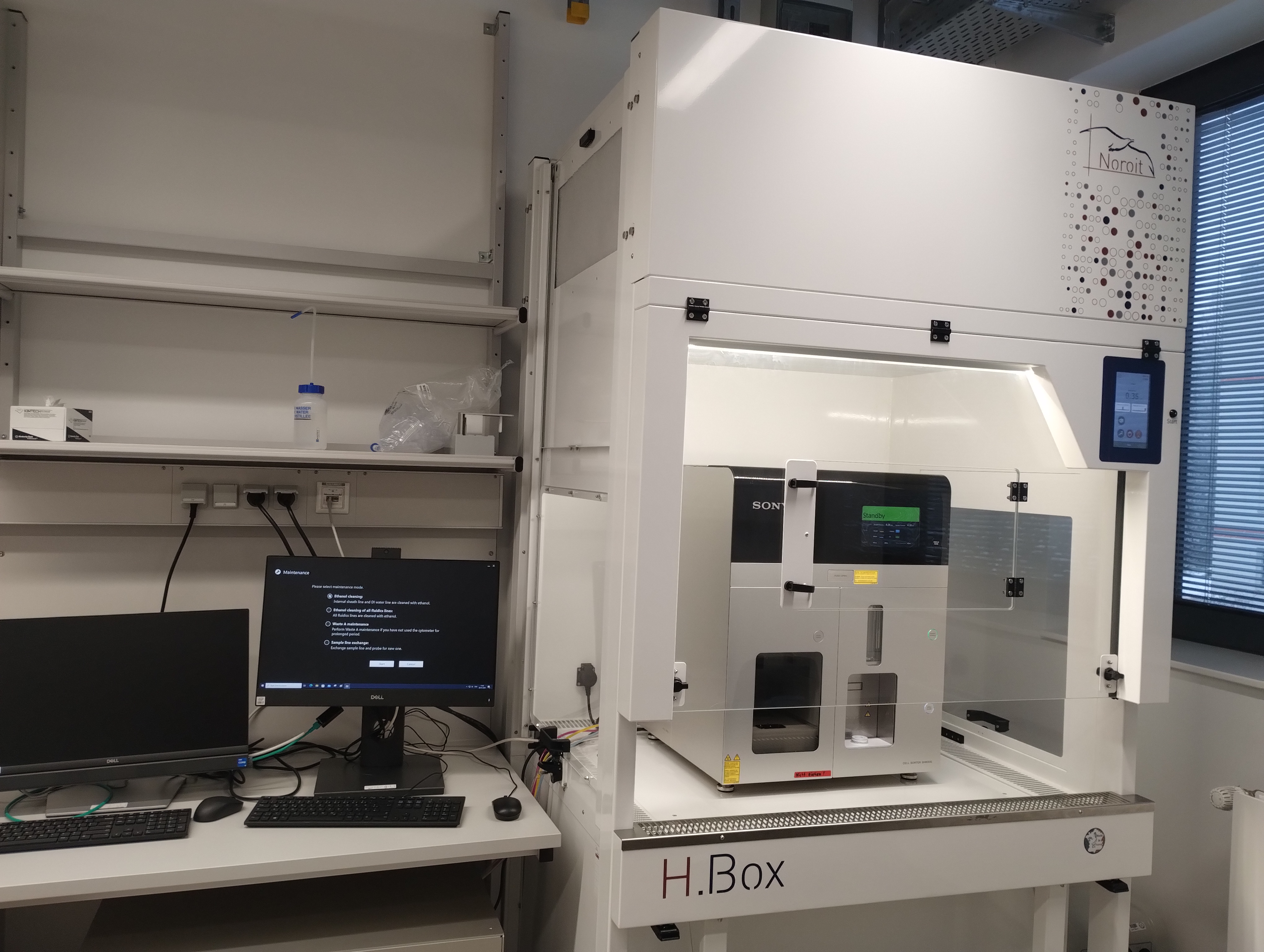
- FACS Sony SH800
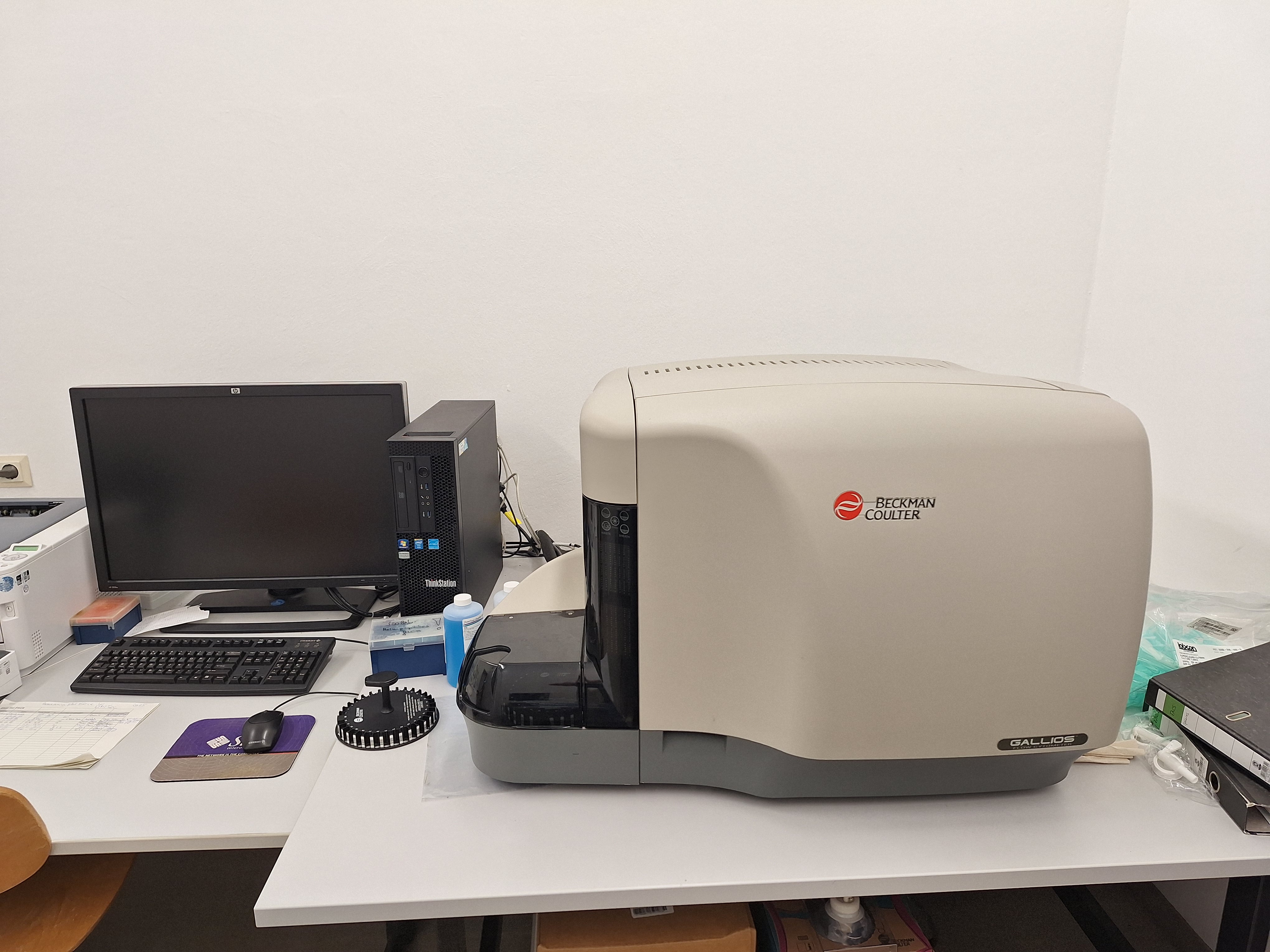
- Beckman Coulter Gallios
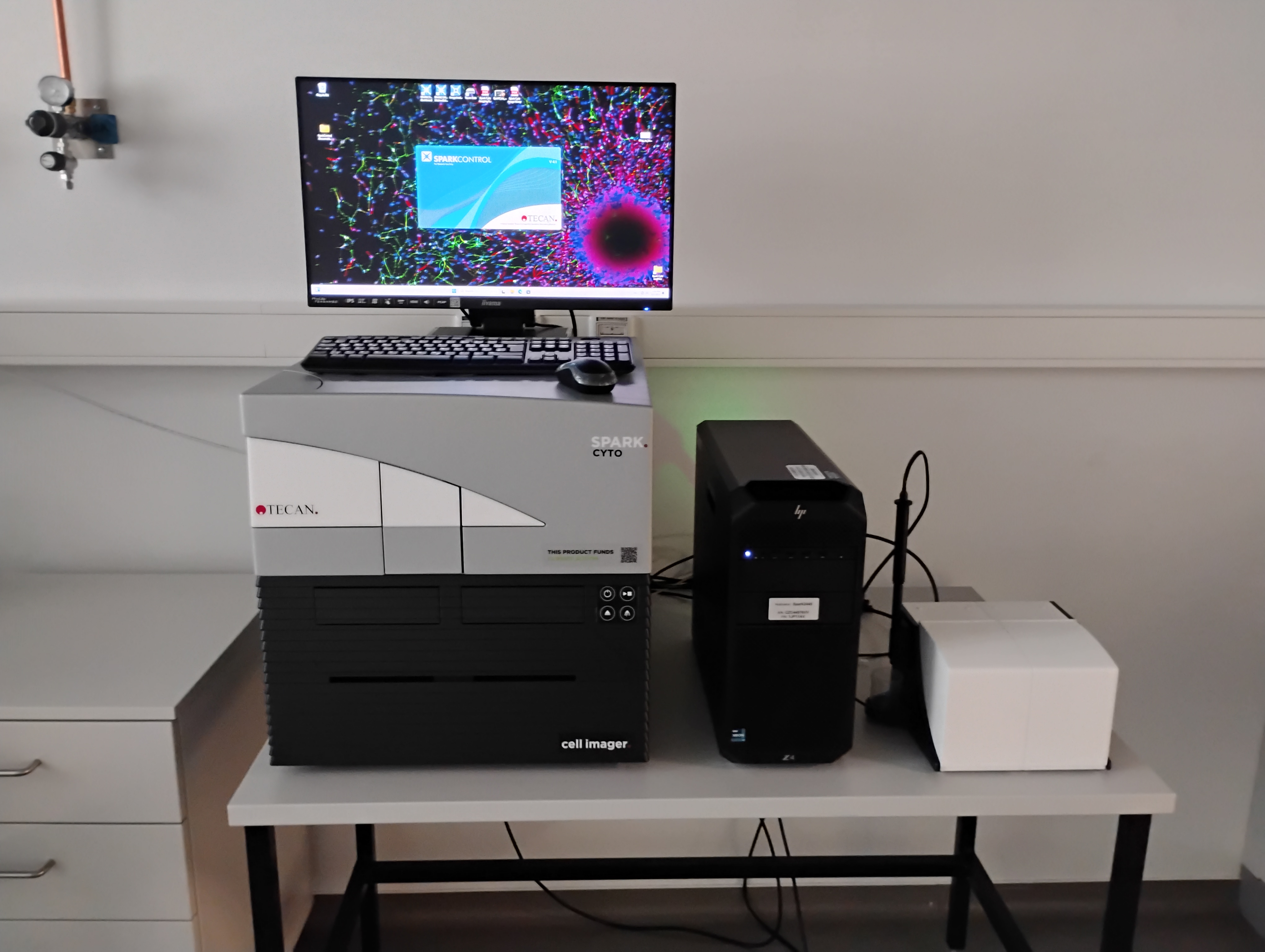
- Tecan Spark Cyto
Multimode Plate Reader (R1 D2-104)
Multimode plate reader with fusion optics (monochromator and filter-based optics) for absorption, polarization, fluorescence modes. Incubation with temperature control and CO2/O2-controller. 2 injectors are available.
Microscopy unit with dry objectives (4x/0.13, 10x/0.30) and filter sets for DAPI, FITC, TRITC and Alexa Fluor 633. Predefined imaging protocols for cell confluency, kinetics, transfection efficiency, nuclei counting, cell vitality.
Contact: Barbara Biermann
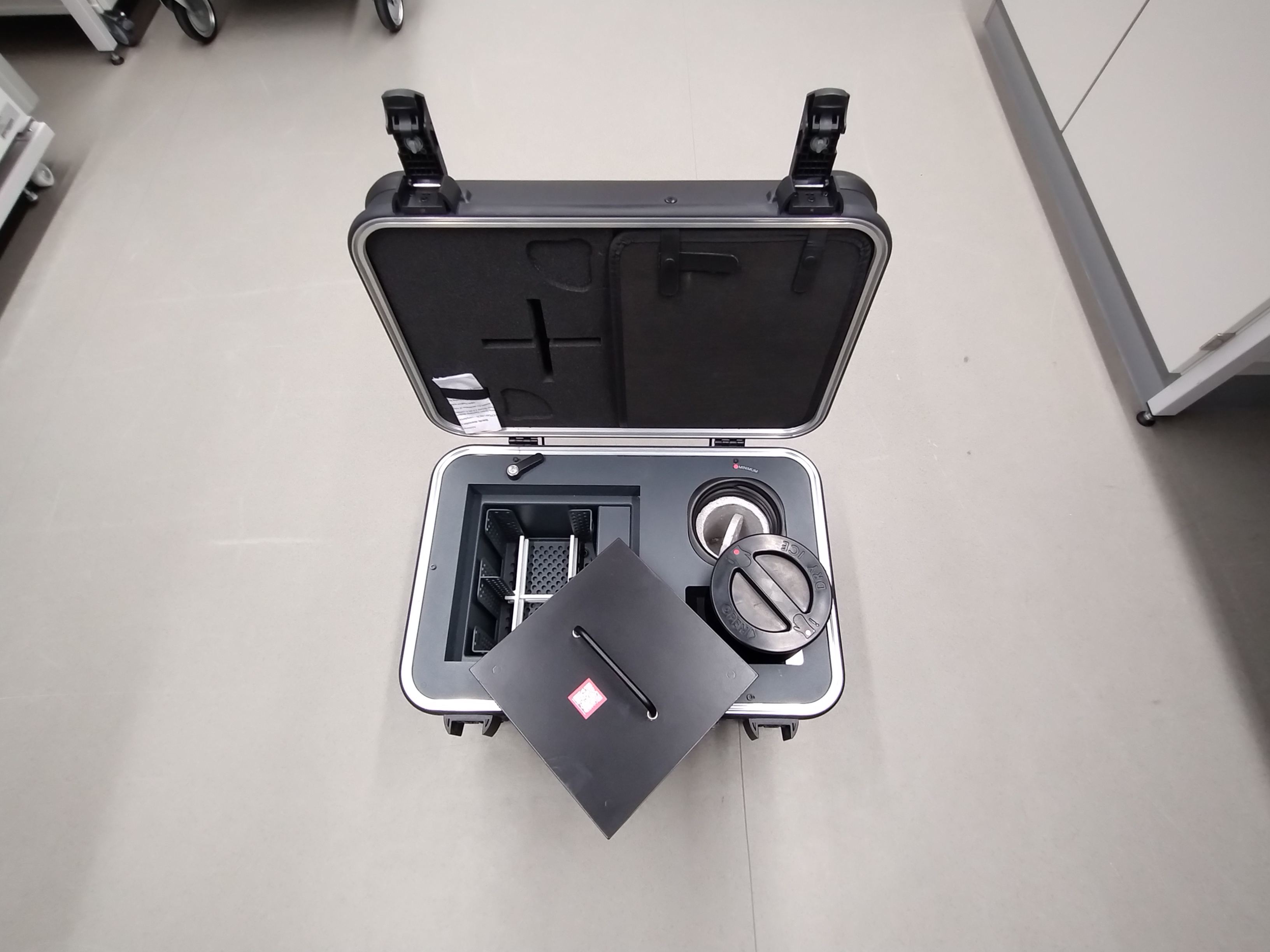
- Cell Box
- Electron Microscopy
We have one transmission electron microscope and two scanning electron microscopes equipped with CCD-cameras.
Sample preparation can be done by our technical staff colleagues, feel free to contact us to schedule the sample submission and discuss the best conditions for your samples.
Contact: tseidel@uni-bielefeld.de
External Instruments
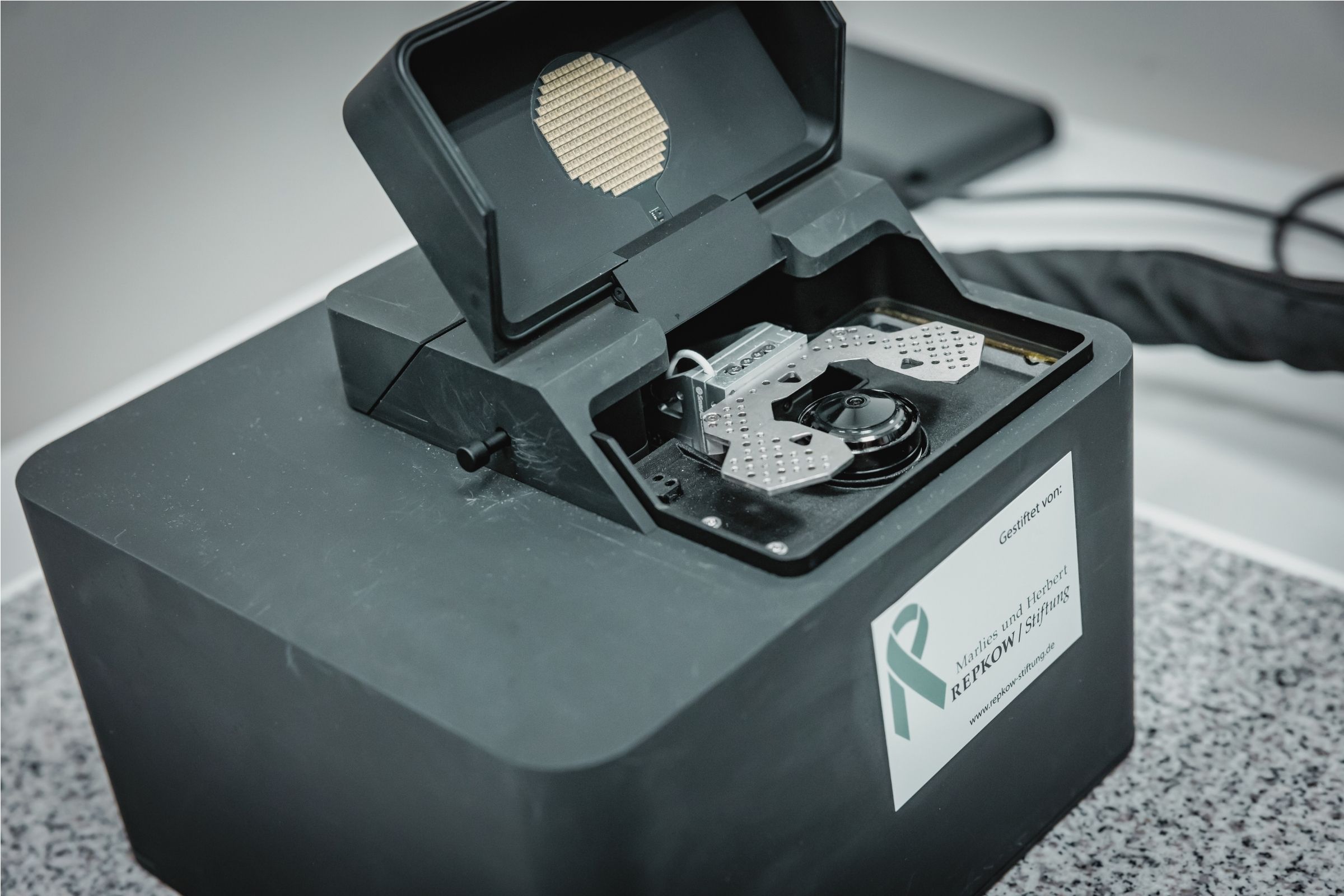
- ONI Nanoimager
Single-molecule localization microscopy (R2, 3. Etage)
- Laser lines: 405 nm, 488 nm, 561 nm, 640 nm
- Objective 100x/1.45
- 2 simultaenous channels (dichroic mirror splitter, 640 nm)
- Hamamatsu Orca Flash4.0 v.3
- lateral resolution of 20 nm
- suitable for single molecule localization microscopy, TIRF and single particle tracking
Prof. Dr. Sven ThomsBiochemie und Molekulare Medizin
Medizinische Fakultät OWL
Phone: 68502Die Anschaffung des Mikroskops war möglich durch eine Sachspende der Marlies und Herbert Repkow Stiftung.
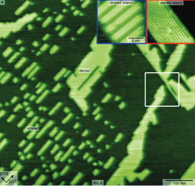
- Atomic Force Microscopy
Omicron STM/AFM
operated in dynamic mode under ultrahigh vacuum conditions
Bruker AFMs (modified)
for atomic-resolution imaging at the solid-liquid interface
solvation layer mapping is possible with home-built hard- and software
Prof. Dr. Angelika KühnlePhysikalische Chemie I
Fakultät für Chemie
Phone: 2045Referenzen:
https://pubs.acs.org/doi/10.1021/acs.jpcc.1c06213
https://doi.org/10.3762/bjnano.11.74
https://doi.org/10.1103/PhysRevB.100.205410
https://doi.org/10.1021/acs.langmuir.6b03814
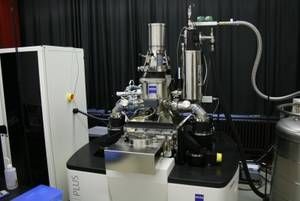
https://doi.org/10.1063/1.4952954- Zeiss Helium-ion Microscope
- Bio Data Mining Group
Field of research / key experience: Development of computational approaches to harvest bioimage data.
Keywords: Bioimage Informatics, Computer Vision, Machine Learning, Remote Sensing, Medical Image Analysis, Information Visualization
Available resource: Online image annotation platform BIIGLE (www.biigle.de) for manual and AI-assisted image / video annotation (development since 2009, > 4,000 users).
Prof. Tim W. Nattkemper (tim@biigle.de)
www.uni-bielefeld.de/fakultaeten/technische-fakultaet/arbeitsgruppen/biodata-miningFaculty of Technology
References
BIIGLE 2.0 - Browsing and Annotating Large Marine Image Collections. Langenkämper D, Zurowietz M, Schoening T, Nattkemper TW. Frontiers in Marine Science, 4, 2017, 83, DOI=10.3389/fmars.2017.00083, ISSN=2296-7745
Current trends and future directions of large scale image and video annotation: Observations from four years of BIIGLE 2.0. M Zurowietz, TW Nattkemper, FRONTIERS IN MARINE SCIENCE (section Ocean Observation), 2021, Manuscript ID: 760036A, https://doi.org/10.3389/fmars.2021.760036