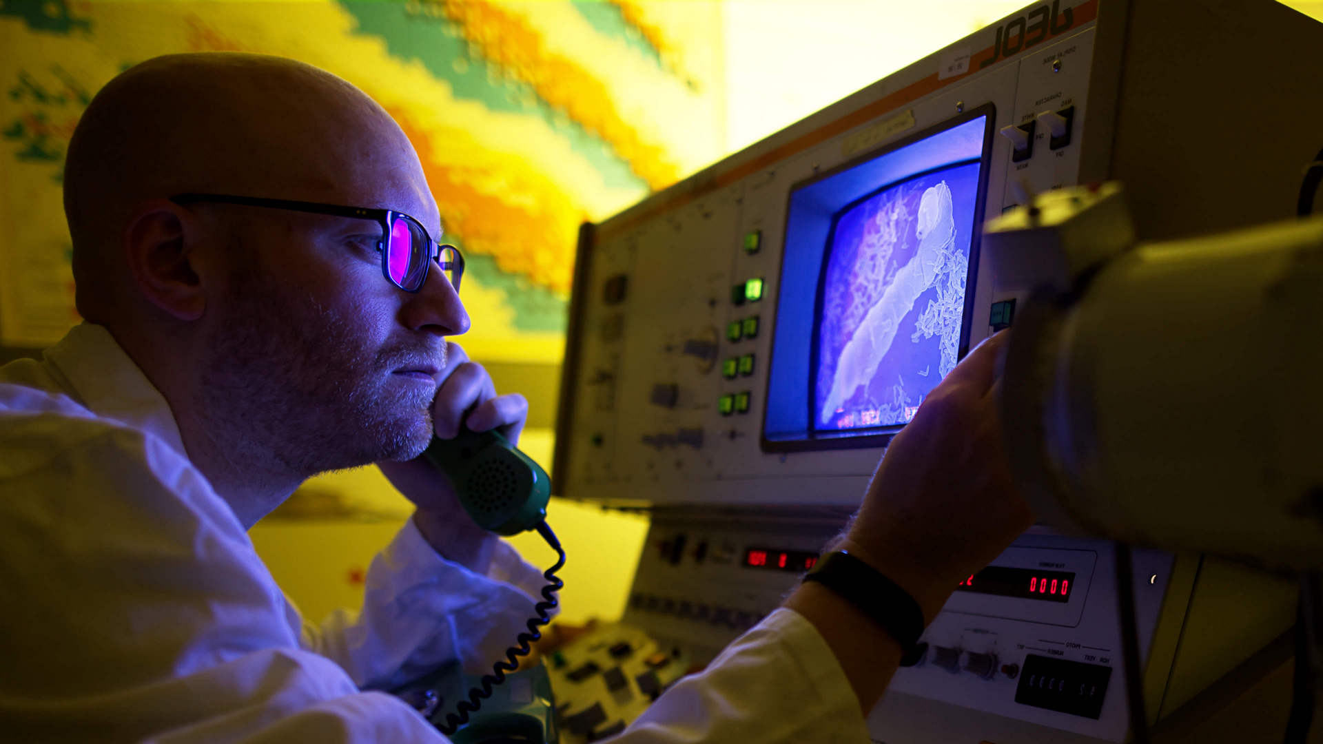
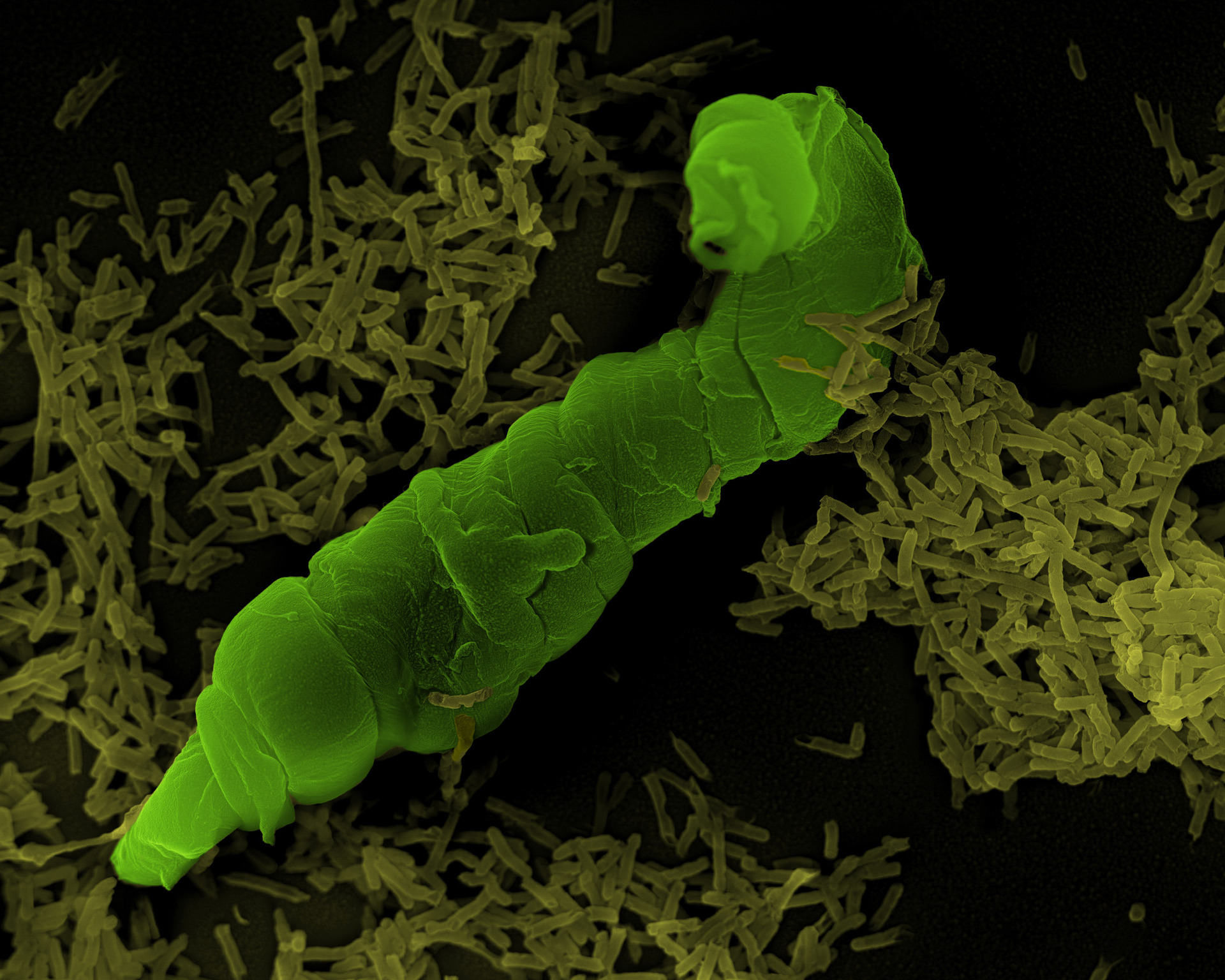
Scanning electron microscopy © Universität Bielefeld/Christoph Pelargus


Scanning probe microscopy
- Atomic Force Microscopy (AFM)
Atomic Force Microscopy (AFM)
STRUCTURAL BIOLOGY
Contact: Dr. Volker Walhorn

Cell of the lung tissue - © Universität Bielefeld/Daniel Wesner Structural information of individual functional molecules and complexes can be investigated by in-situ atomic force microscopy (AFM) by immobilising the biomolecules of interest (nucleic acids, proteins, carbohydrates, …) via physical, chemical or biological interaction on a flat surface or directly embedded in the cellular membrane environment. The interesting structural information includes sub-nm-conformation, molecular symmetry, binding location and molecular temporal dynamics. The following typical examples are typical results of actual research projects where biological processes of higher complexity (transcription regulation,molecular motors, self-assembly of 2D-protein s-layers) are investigated.
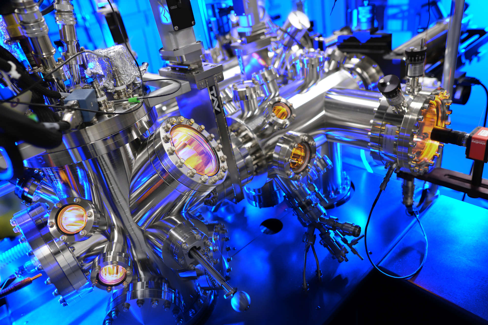
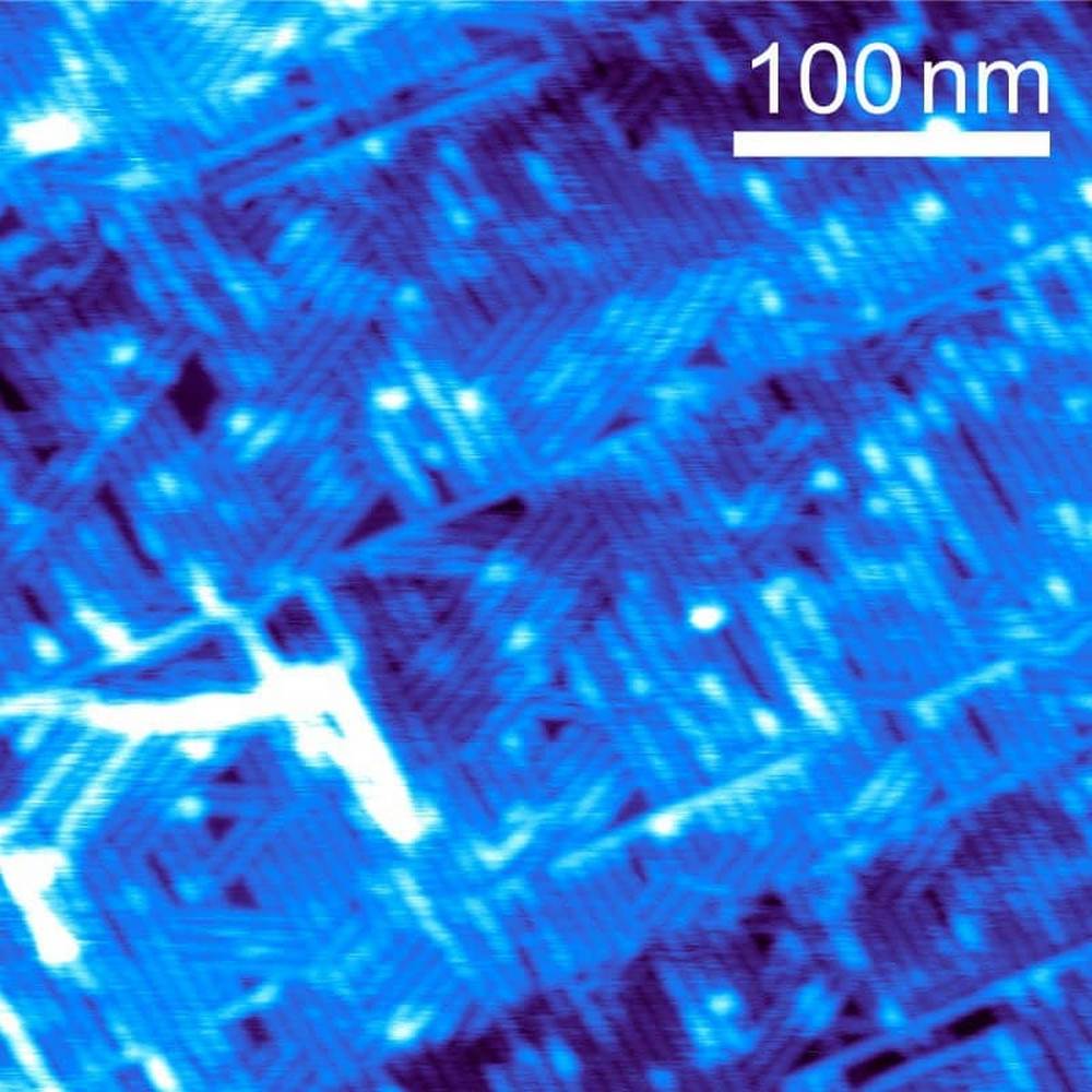
We currently run several commercial AFM for experiments under ambient and liquid conditions, self-built combined TIRF/AFM and SNOM microscopes, as very recently a novel STM/AFM-microscope for ultra-high-vacuum applications.© Universität Bielefeld/Fabian Heising Diyne-phosphocholine molecules arrange themselves after Langmuir-Blodgett transfer onto Highly Ordered Pyrolytic Graphite - a step towards simulating bilipid cell membranes.
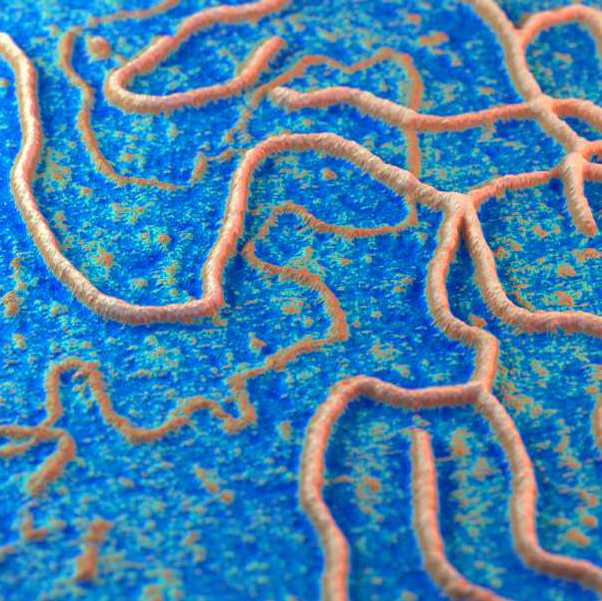
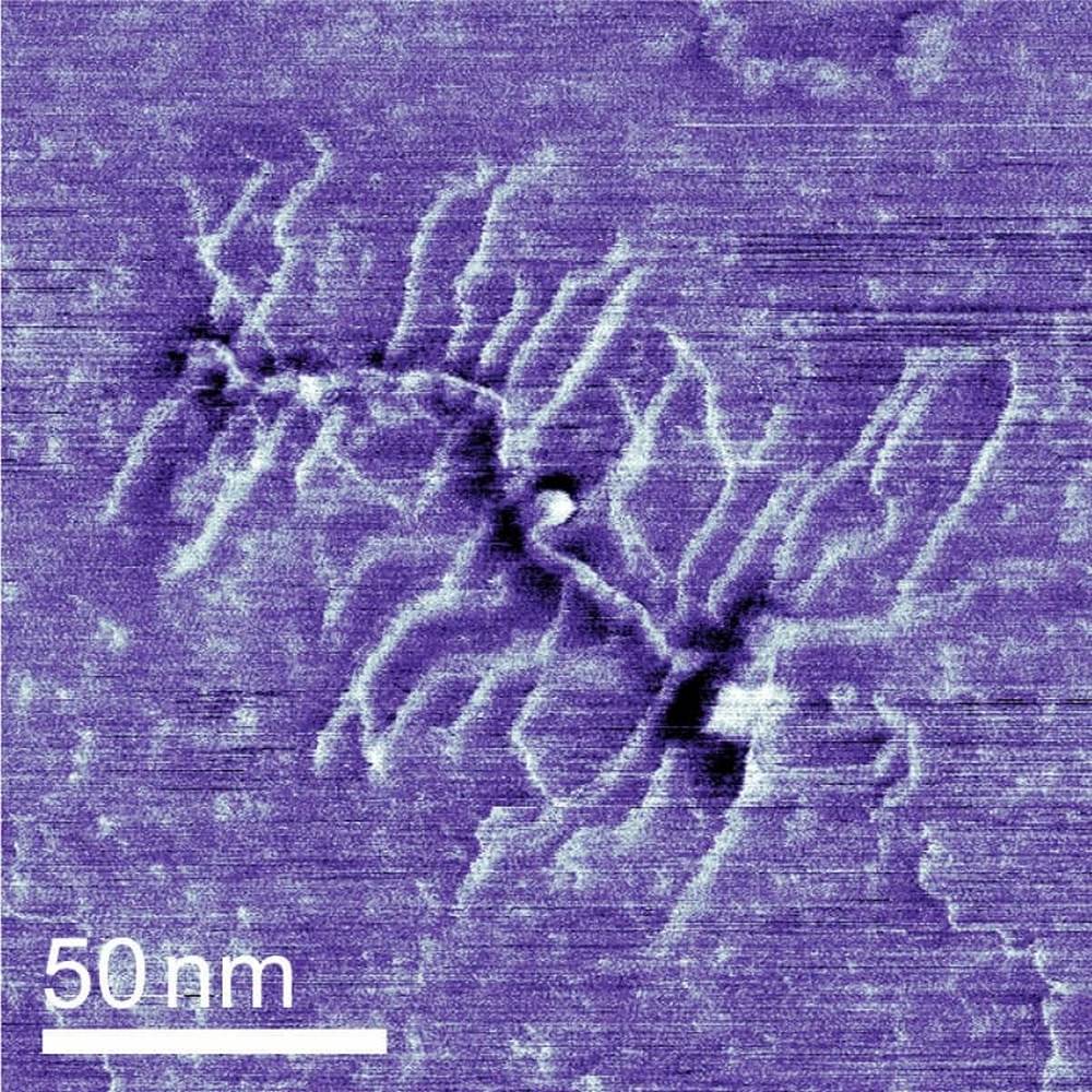
© Universität Bielefeld/Niklas Biere A proteoglycan molecule from the sea cucumber Isostichopus badionotus has a brush-like structure. The delicate arms can bind water and this way change the mechanical properties of their tissue.
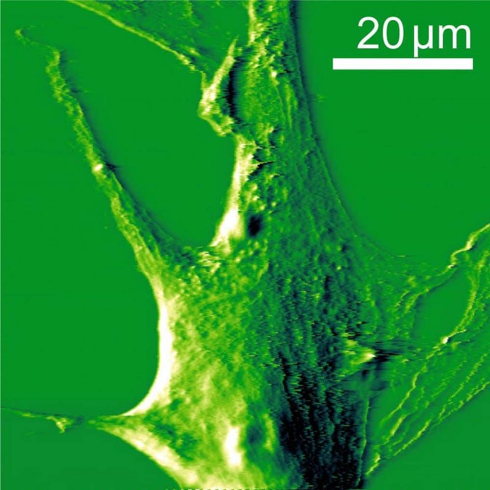
© Universität Bielefeld/Orooba Al-Hammood This fibroblast cell from human tissue is fixated and prepared for force spectroscopic measurements - to determine its elastic properties and ultimately their relation to genetic heart diseases.
- Single Molecule AFM Force Spectroscopy
Single Molecule AFM Force Spectroscopy
PHYSICS OF MOLECULAR RECOGNITION
Contact: Dr. Volker Walhorn
Flowbox workbench - © Universität Bielefeld/Niklas Biere The physical mechanisms of specific, non-covalent intermolecular binding is quantitatively investigated by single-molecule (dynamic) force spectroscopy with atomic force microscopy (AFM), optical tweezers (OT) and magnetic tweezers (MT). Here, specific molecular forces, elasticities, kinetic reaction rate constants (lifetimes) and the energy landscape of the molecular binding potential is measured by AFM at the single-molecule level in vitro on isolated but functional complexes. Nowadays, these specific interactions can be scrutinized in a quantitative manner at the sensitivity level on single-point mutations (nucleic acids, amino acids) allowing single-molecule affinity ranking in a broad affinity range of 0,1 mM-1 fM revealing distinct differences in the binding properties and mechanisms. Over the last years, special emphasis has been put on so-called molecular catch-bond systems that were observed when investigating the interplay of human sulfatates (Sulf1, Sulf2) with glycosaminoglycans (heparan sulfate, heparin, dermatan sulfate, ...) in cell signaling cascades. In addition, we recently explored the homophilic interaction mechanisms between desmosomal desmoglein-2 (DSG2) and mutations thereof that play an important role for the integrity of mechanically active cells like cardiomyocytes.
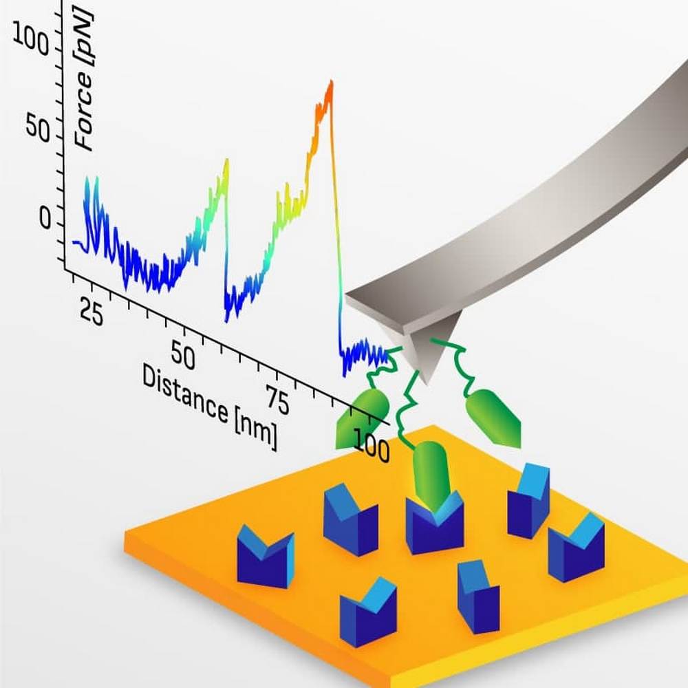
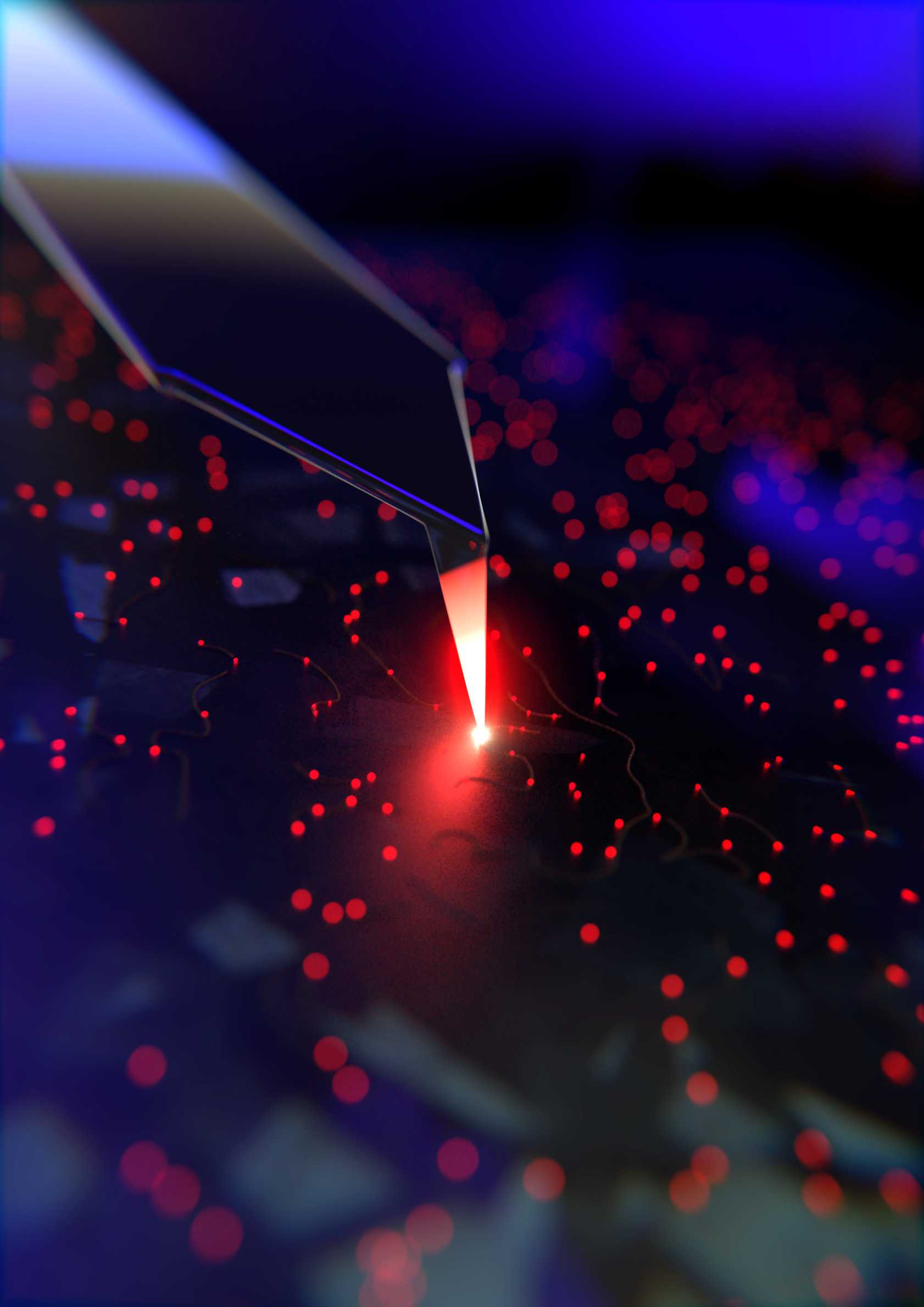
© Universität Bielefeld/Niklas Biere Tip and sample surface are both functionalized. In close proximity, individual molecular bonds are formed. When pulled apart, ruptures can be seen as a jumps in the force curve. The corresponding binding forces and lifetimes can be measured with high precision, giving insight in biological relevant binding and sensing processes and even protein folding.
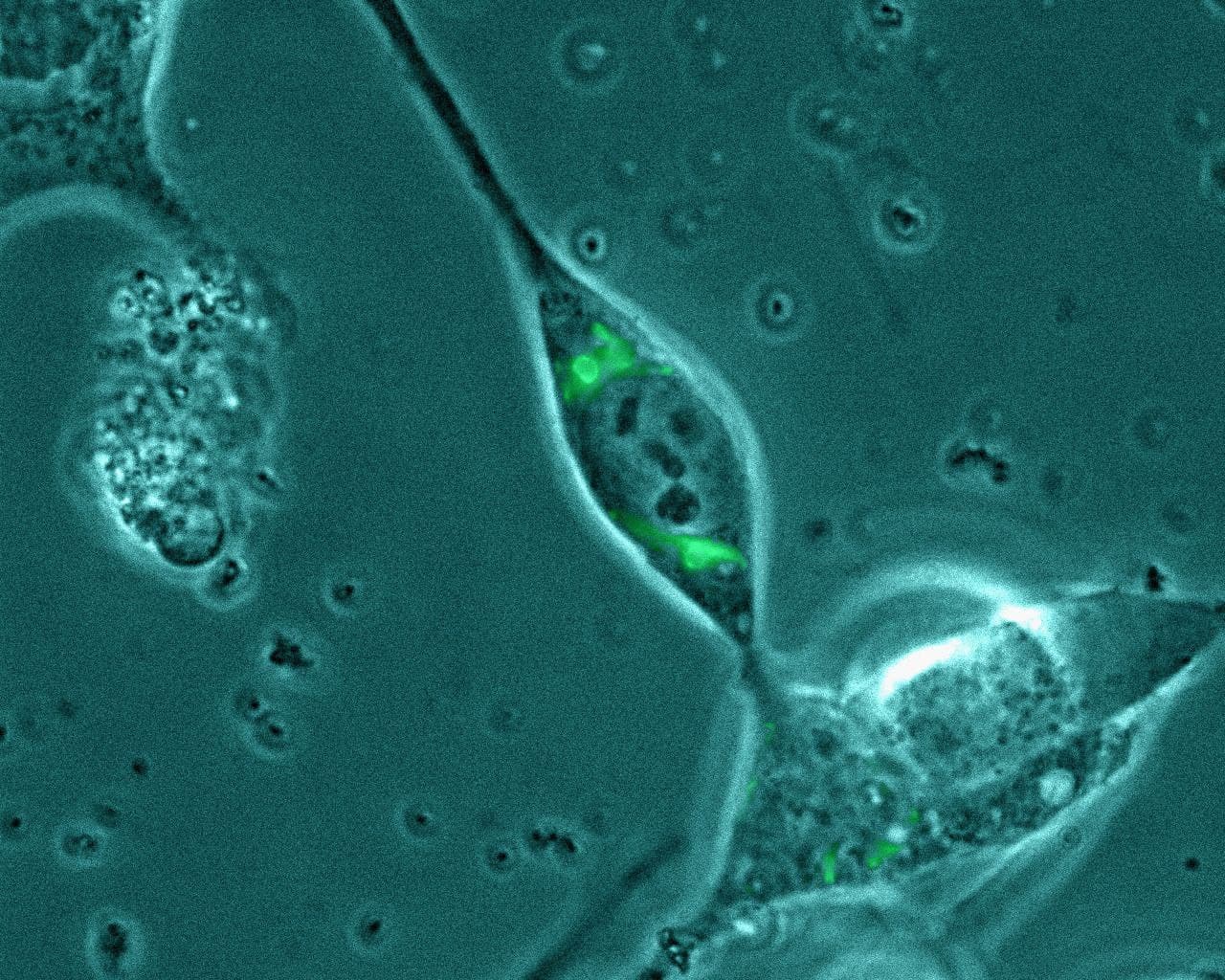
© Universität Bielefeld/Esther Borchers Fluorescent labeled cell compartments help guide where to poke the cell - to measure the elasticity in regards to specific organelles that might be affected by genetic conditions.
- Single Molecule Photonics
Single Molecule Photonics
AFM-TIRF / SNOM
Contact: Dr. Volker Walhorn
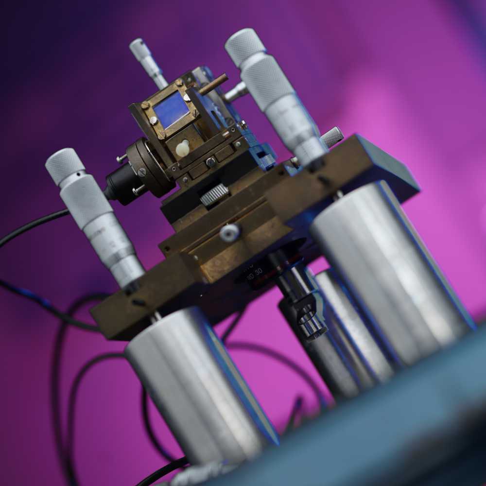
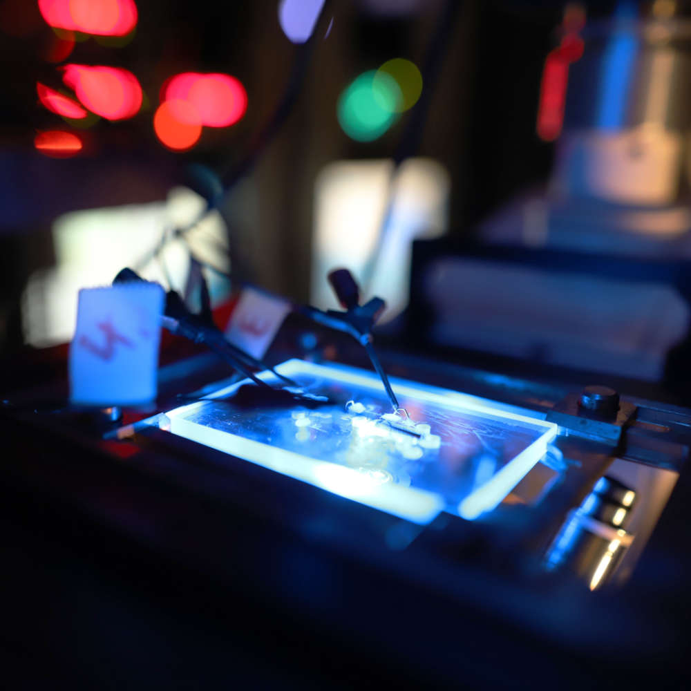
Self made TIRF combined AFM head - © Universität Bielefeld/C.Pelargus Single molecules can nowadays be investigated by means of optical, mechanical and electrical methods. Single molecule fluorescence imaging and spectroscopy yield valuable and quantitative information about optical properties, spatial distribution and temporal dynamics of single molecules.
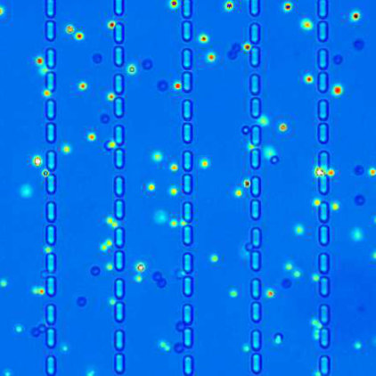
We have developped 1) scanning near-field optical microscopy (SNOM) for single molecule imaging and spectroscopy and 2) a small-cantilever based AFM combined with total internal reflection fluorescence (TIRF) microscopy for ultrasensitive LIF detection of individual fluorophores and simultaneous AFM control and manipulation at the molecular level.© Universität Bielefeld/Niklas Biere Individual fluorescent markers can be localized in the AFM topography with super resolution precision - here shown in tobacco mosaic viruses.
© Universität Bielefeld/Volker Walhorn Simulation of an electric field at a gold tip 5nm above an air-glass interface. The configuration is illuminated from below at an angle of 50°, so that an evanescent field is formed above the interface
© Universität Bielefeld/Niklas Biere Evanescent illumination causes several hundred-times higher field intensities at the cantilever apex as usual - also enhancing the radiative emission of fluorophor


Nanomanipulation
- Nanopores
Nanopores
SINGLE MOLECULE TRANSLOCATION
Contact: Prof. Dr. Dario Anselmetti
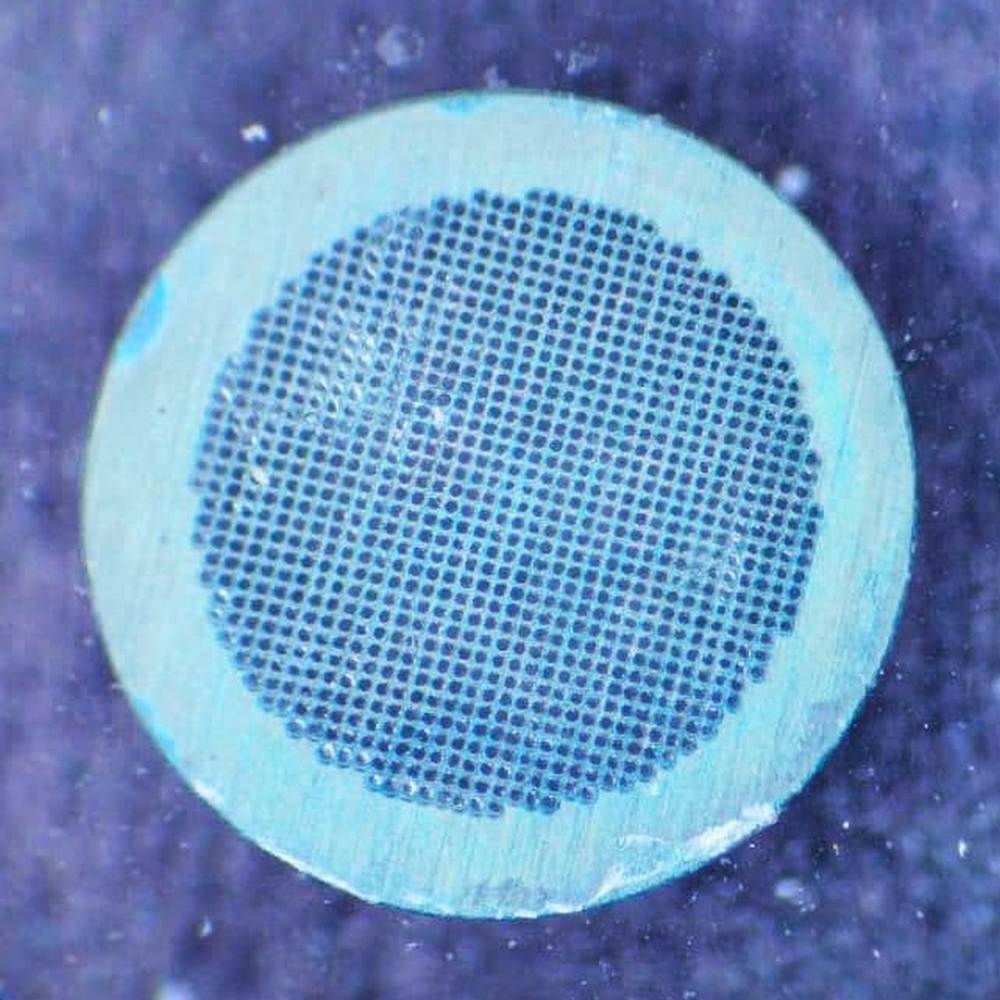
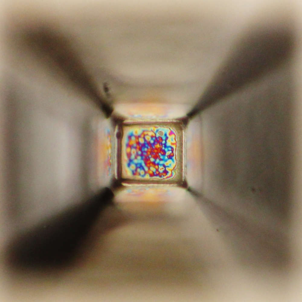
SEM grid for preparation - © Universität Bielefeld/Roland Hillmann The translocation dynamics of individual macromolecules like DNA or DNA-protein complexes (EcoRI, RecA, peroxiredoxines) through solid-state nanopores (SiNx, graphene, MoS2, BN, ...) is quantitativly investigated by optical tweezers force control. Beyond looking into the physical mechanisms involved we explore the possibility of future sequencing and diagnostic applications thereof.
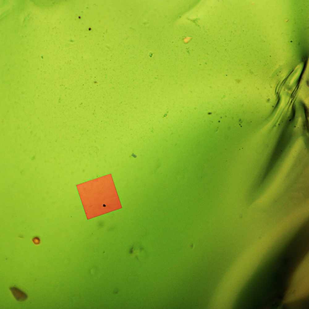
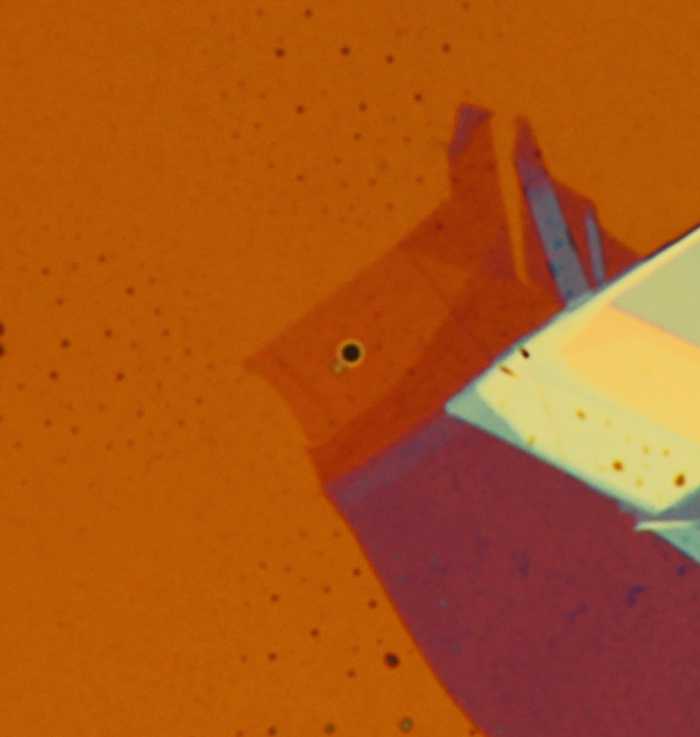
© Universität Bielefeld/Dennis Kreft Pore cover from backside ,optical microscope view, while evaporating the solvent.
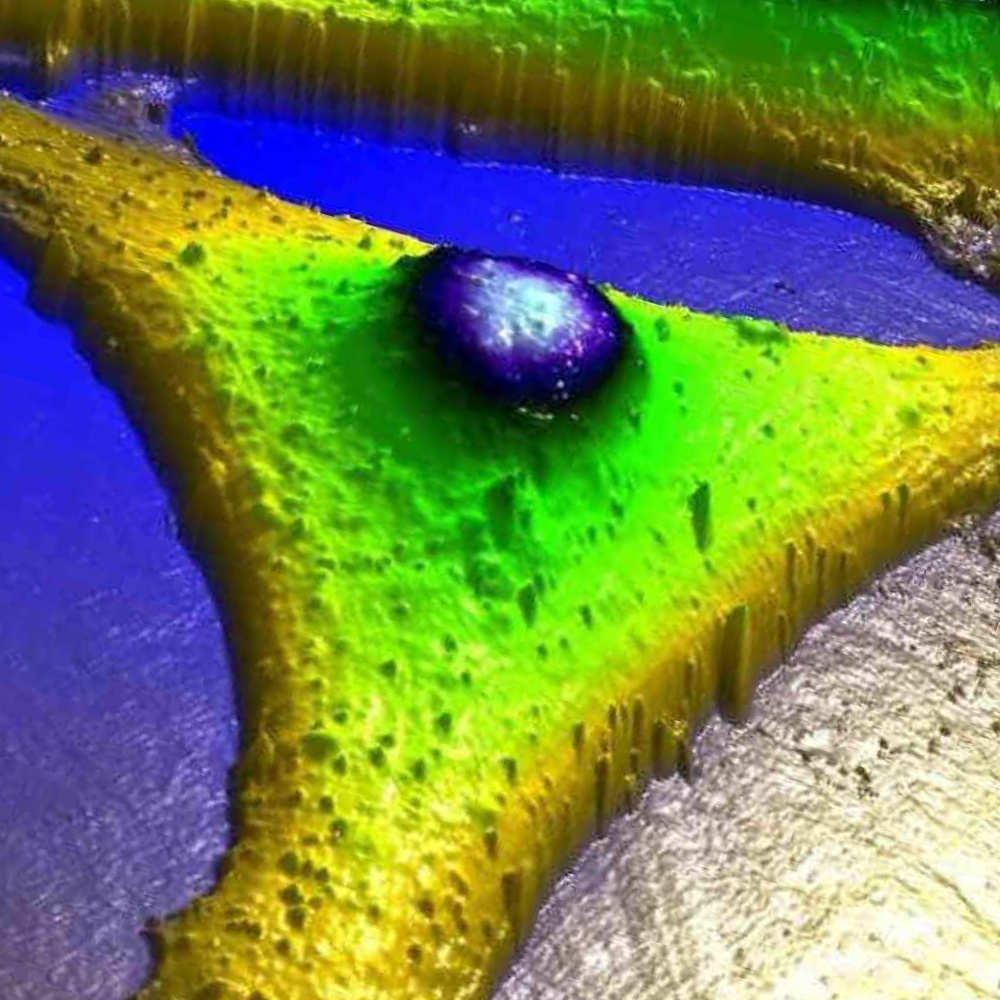
© Universität Bielefeld/Julian Cremer Freestanding MoS2-monolayer with a thickness of 0.67nm covering a 600nm opening
- Optical Tweezers
Optical Tweezers
SINGLE MOLECULE BIOSENSOR & MANIPULATION
Contact: Prof. Dr. Dario Anselmetti
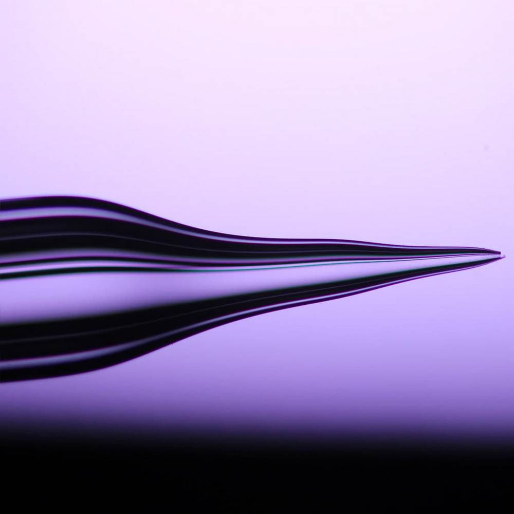
Glass capillary - © Universität Bielefeld/Tobias Jäckering Micron-sized objects like beads, colloids or cells can be trapped, steered and manipulated by light and allow force experiments at the single molecule level with a force sensitivity level of 0.1 pN. We set up two high stability single-beam optical tweezers (OT) system on an inverted light microscope which allows analytical force spectroscopy, adhesion and elasticity experiments with single molecules or cells in an experimental force range of 0.1-900 pN.
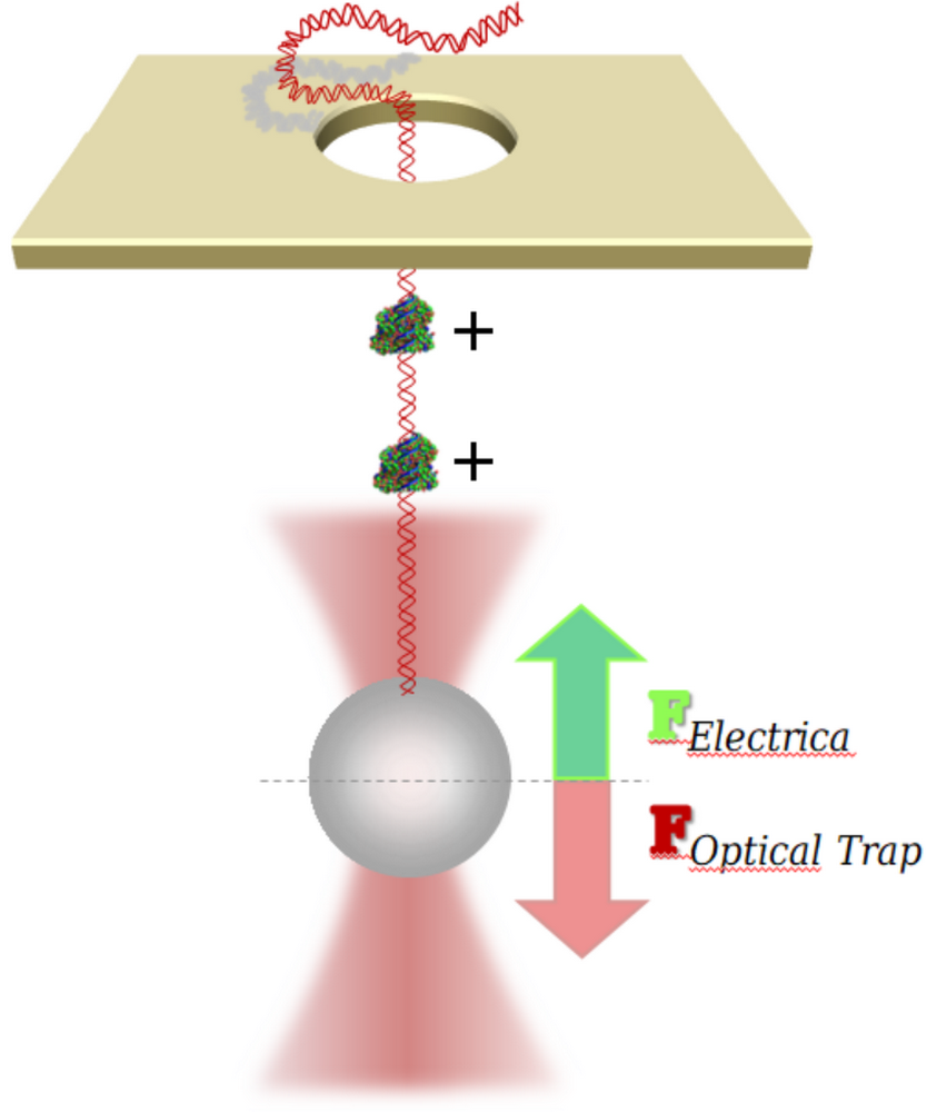
© Universität Bielefeld/Lukas Galla Principle in the experiments with controlled DNA translocations through nanopores with optical tweezers.
- Magnetic Tweezers
Magnetic Tweezers
SINGLE MOLECULE MANIPULATION WITH MAGNETIC FIELDS
Contact: Prof. Dr. Dario Anselmetti
Different buffer concentrations © Universität Bielefeld/Niklas Biere Magnetic microbeads can be manipulated and steered by external magnetic fields and allow stretching and overwinding experiments with single (DNA) molecules at a sensitivity level downto 0,001 pN. With our magnetic tweezers (MT) apparatus (PicoTwist, Lyon - France) we investigate and quantify the binding of proteins, fluorescent dyes and chemotherapeutic agents to DNA.


Microfluidics
- Microfluidics
Micro- & Nanofluidics
MIGRATION & DYNAMICS OF BIOMOLECULES
Contact: Dr. Martina Viefhues
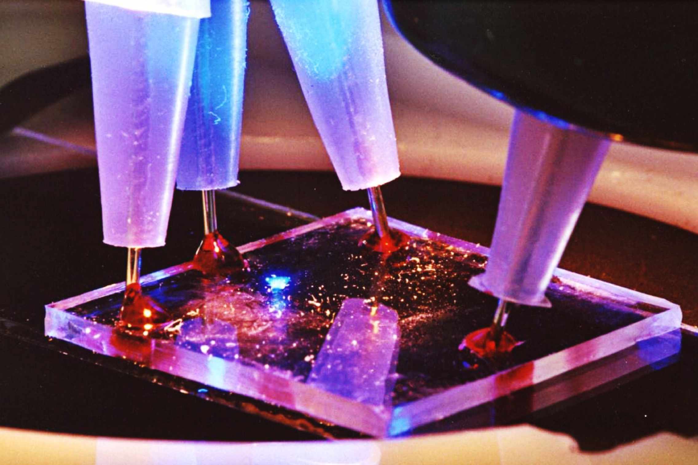
PDMS chip with electrodes - © Universität Bielefeld/C.Pelargus In this research project we investigate and develop novel micro- and nanofluidical chip device applications that can be used for separation, sorting and harvesting of macromolecules and colloids. We use structured micro- and nanofluidic environments and drive the molecular objects far from thermoddynamic equilibrium to explore non-linear migration mechanisms like absolute negative mobility, dielectrophoretic trapping, molecular ratcheting or chiral separation.
In addition, we design and develop lab-on-a-chip devices where individual cells like E. coli can be trapped and analyzed according their active metabolism by UV-laser-induced fluorescence.© Universität Bielefeld/Armin Grundmann Microscopy picture of a 20µm width PDMS microchanel crossing with a cell trap.
© Universität Bielefeld/Christoph Pelargus Microfluidic flow cell with high voltage electrodes conducted and
fluorescence detection.

Tribologie
- Industrial Projects (BINAS)
Industrial Projects (BINAS)
NANOPARTICLES, TRIBOLAB, ...
Contact: Dr. Katja Tönsing
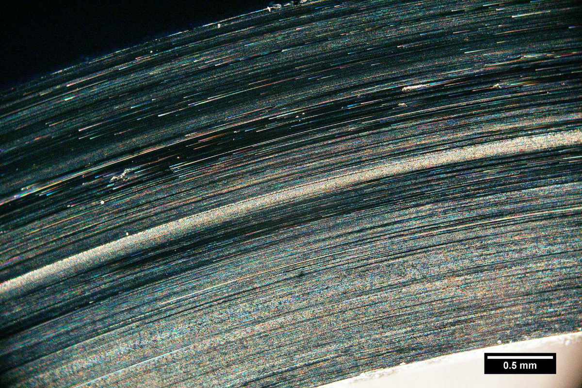
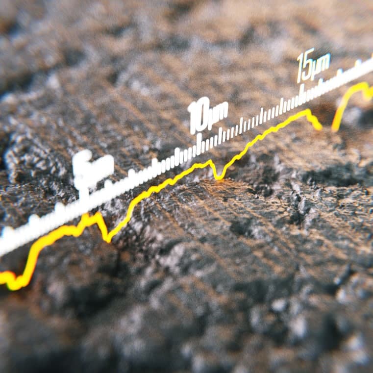
Test disc with worn surface - © Universität Bielefeld/Alexander Beel In this research and development project we investigate and develop new methods and products for use in industrial applications. Together with our industrial partners we analyze, develop and optimize nanotechnological products and processes ranging from e.g. nanoparticle based lubricating performance systems, nanomembrane-based biosensor surfaces as well as effective microchip processes for separating large genomic mixtures.
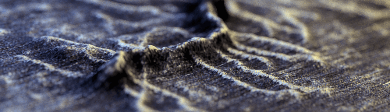
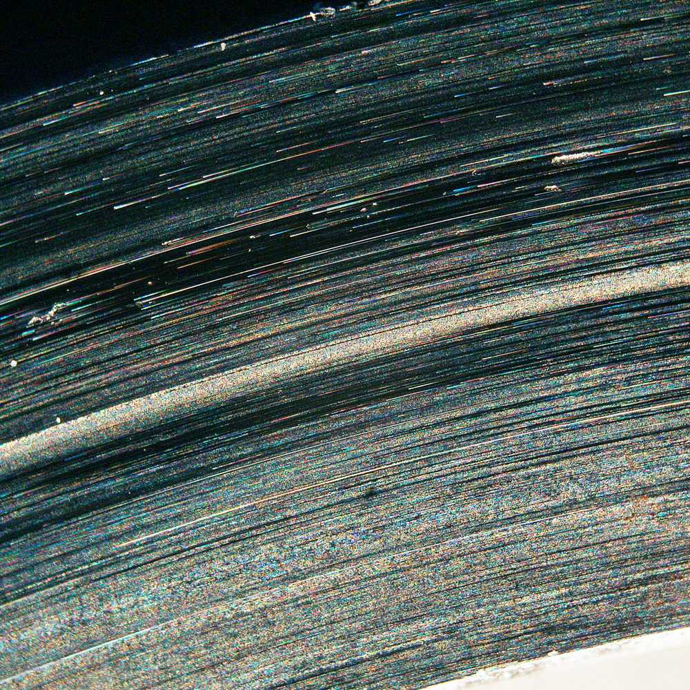
© Universität Bielefeld/Alexander Beel/Niklas Biere Surface of a grey cast iron disk.
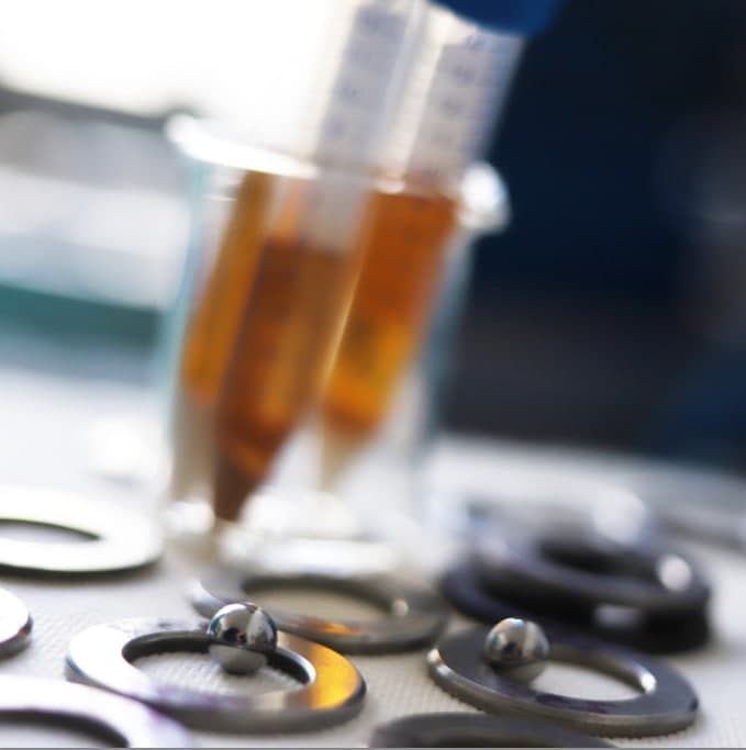
© Universität Bielefeld/Niklas Biere Test samples with marbles made of hardened steel for friction measurement. Different oil mixtures can be testet.
Diverses
- Job offers
Thema Ansprechpartener Infos Bachelorarbeiten / Abschlussarbeiten fürs Lehramt ... nach Absprache Prof. Dario Anselmetti (D1-270) Kurzbeschreibung (.pdf) Masterarbeiten
... nach Absprache Prof. Dario Anselmetti (D1-270) Kurzbeschreibung (.pdf) Doktorarbeiten / Ph.D. Thesis
... nach Absprache Prof. Dario Anselmetti (D1-270) Kurzbeschreibung (.pdf) - Coperations
Currently, we acknowledge funding from the following research grants:
- German Science Foundation (DFG) - DA 370/6-1: Molecular mechanisms of Desmin-ARVC-cardiomyopathy (2015-2017)
- German Science Foundation (DFG) - DA 370/7-1: Translocation through biological nanopores (2015-2017)
- German Science Foundation (DFG) - DA 370/8-1: Dinuclear complexes that bind to DNA (2016-2019)
- Industry project (Evonik): Nanoparticles and nanofluids (2016-2018)
- EU-Rocket Interreg Germany / Netherlands: Elasto-Tweezers (2017-2019)
- Forschungsfonds Medizin NRW/RUB/UniBi: Investigation of TMEM43-related cardiomyopathies (2017-2019)